 Movie
Movie Controller

[English] 日本語
 Yorodumi
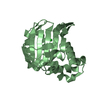
- PDB-1rbz: Human GAR Tfase complex structure with polyglutamated 10-(trifluo... -
+ Open data

- Basic information
| Entry | Database: PDB / ID: 1rbz | ||||||
|---|---|---|---|---|---|---|---|
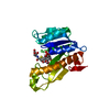
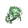
| Title | Human GAR Tfase complex structure with polyglutamated 10-(trifluoroacetyl)-5,10-dideazaacyclic-5,6,7,8-tetrahydrofolic acid | ||||||
 Components Components | PHOSPHORIBOSYLGLYCINAMIDE FORMYLTRANSFERASE | ||||||
 Keywords Keywords | TRANSFERASE / PROTEIN-COFACTOR ANALOGUE COMPLEX | ||||||
| Function / homology |  Function and homology information Function and homology informationphosphoribosylamine-glycine ligase / phosphoribosylglycinamide formyltransferase 1 / adenine biosynthetic process / phosphoribosylformylglycinamidine cyclo-ligase / phosphoribosylformylglycinamidine cyclo-ligase activity / phosphoribosylglycinamide formyltransferase activity / purine ribonucleoside monophosphate biosynthetic process / phosphoribosylamine-glycine ligase activity / brainstem development / 'de novo' XMP biosynthetic process ...phosphoribosylamine-glycine ligase / phosphoribosylglycinamide formyltransferase 1 / adenine biosynthetic process / phosphoribosylformylglycinamidine cyclo-ligase / phosphoribosylformylglycinamidine cyclo-ligase activity / phosphoribosylglycinamide formyltransferase activity / purine ribonucleoside monophosphate biosynthetic process / phosphoribosylamine-glycine ligase activity / brainstem development / 'de novo' XMP biosynthetic process / Purine ribonucleoside monophosphate biosynthesis / 'de novo' AMP biosynthetic process / purine nucleotide biosynthetic process / GMP biosynthetic process / 'de novo' IMP biosynthetic process / cerebellum development / cerebral cortex development / extracellular exosome / ATP binding / metal ion binding / cytosol Similarity search - Function | ||||||
| Biological species |  Homo sapiens (human) Homo sapiens (human) | ||||||
| Method |  X-RAY DIFFRACTION / SYNCHROTRON / MOLECULAR REPLACEMENT / Resolution: 2.1 Å X-RAY DIFFRACTION / SYNCHROTRON / MOLECULAR REPLACEMENT / Resolution: 2.1 Å | ||||||
 Authors Authors | Zhang, Y. / Desharnais, J. / Boger, D.L. / Wilson, I.A. | ||||||
 Citation Citation | Journal: To be Published Title: Human GAR Tfase complex structure Authors: Zhang, Y. / Desharnais, J. / Boger, D.L. / Wilson, I.A. | ||||||
| History |
|
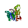
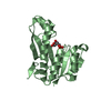
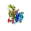
- Structure visualization
| Structure viewer | Molecule: MolmilJmol/JSmol |
|---|
- Downloads & links
-Download
| PDBx/mmCIF format | 1rbz.cif.gz | 93 KB | Display | PDBx/mmCIF format |
|---|---|---|---|---|
| PDB format | pdb1rbz.ent.gz | 71.1 KB | Display | PDB format |
| PDBx/mmJSON format | 1rbz.json.gz | Tree view | PDBx/mmJSON format | |
| Others |  Other downloads Other downloads |
-Validation report
| Arichive directory | https://data.pdbj.org/pub/pdb/validation_reports/rb/1rbzftp://data.pdbj.org/pub/pdb/validation_reports/rb/1rbz | HTTPS FTP |
|---|
-Related structure data
| Related structure data |  1rbmC  1rbyC  1rc0C  1rc1C  1njsS S: Starting model for refinement C: citing same article ( |
|---|---|
| Similar structure data |


-Links
 PDBj
PDBj


- Assembly
| Deposited unit | 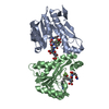
| ||||||||||
|---|---|---|---|---|---|---|---|---|---|---|---|
| 1 | 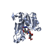
| ||||||||||
| 2 | 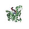
| ||||||||||
| Unit cell |
|
-Components
| #1: Protein | Mass: 22678.941 Da / Num. of mol.: 2 / Fragment: (residues 808-1010) Source method: isolated from a genetically manipulated source Details: part of Trifunctional purine biosynthetic protein adenosine-3 Source: (gene. exp.) Homo sapiens (human) / Gene: purN / Plasmid: pet22a / Production host:  References: UniProt: P22102, phosphoribosylglycinamide formyltransferase 1 #2: Chemical | 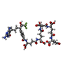  Mass: 1061.922 Da / Num. of mol.: 2 / Source method: obtained synthetically / Formula: C42H54F3N9O20 Mass: 1061.922 Da / Num. of mol.: 2 / Source method: obtained synthetically / Formula: C42H54F3N9O20#3: Water | ChemComp-HOH / |  Mass: 18.015 Da / Num. of mol.: 153 / Source method: isolated from a natural source / Formula: H2O Mass: 18.015 Da / Num. of mol.: 153 / Source method: isolated from a natural source / Formula: H2O |
|---|
-Experimental details
-Experiment
| Experiment | Method: X-RAY DIFFRACTION / Number of used crystals: 1 |
|---|
- Sample preparation
| Crystal | Density Matthews: 4.71 Å3/Da / Density % sol: 73.7 % |
|---|---|
| Crystal grow | Temperature: 277 K / Method: vapor diffusion, sitting drop / pH: 4 Details: PEG 1500, Sodium Acetate, pH 4., VAPOR DIFFUSION, SITTING DROP, temperature 277K |
-Data collection
| Diffraction | Mean temperature: 100 K |
|---|---|
| Diffraction source | Source: SYNCHROTRON / Site: SSRL  / Beamline: BL11-1 / Wavelength: 0.992 Å / Beamline: BL11-1 / Wavelength: 0.992 Å |
| Detector | Type: ADSC QUANTUM 9 / Detector: CCD / Date: Jun 14, 2002 / Details: mirrors |
| Radiation | Monochromator: GRAPHITE / Protocol: SINGLE WAVELENGTH / Monochromatic (M) / Laue (L): M / Scattering type: x-ray |
| Radiation wavelength | Wavelength: 0.992 Å / Relative weight: 1 |
| Reflection | Resolution: 2.06→47 Å / Num. all: 53508 / Num. obs: 53508 / % possible obs: 99.3 % / Observed criterion σ(I): -3 / Redundancy: 4.15 % / Rsym value: 0.07 / Net I/σ(I): 24.9 |
| Reflection shell | Resolution: 2.06→2.13 Å / Redundancy: 3.68 % / Mean I/σ(I) obs: 1.71 / Num. unique all: 5186 / Rsym value: 0.614 / % possible all: 96.8 |
- Processing
| Software |
| |||||||||||||||||||||||||
|---|---|---|---|---|---|---|---|---|---|---|---|---|---|---|---|---|---|---|---|---|---|---|---|---|---|---|
| Refinement | Method to determine structure: MOLECULAR REPLACEMENT Starting model: 1NJS Resolution: 2.1→47 Å / Cross valid method: THROUGHOUT / σ(F): 0 / Stereochemistry target values: Engh & Huber
| |||||||||||||||||||||||||
| Displacement parameters |
| |||||||||||||||||||||||||
| Refinement step | Cycle: LAST / Resolution: 2.1→47 Å
| |||||||||||||||||||||||||
| Refine LS restraints |
|
