[English] 日本語
Yorodumi
- PDB-5j9f: Human GAR transformylase in complex with GAR and (4-{[2-(2-Amino-... -

+
Open data


ID or keywords:

Loading...

-
Basic information

Entry
Database: PDB / ID: 5j9f
Title
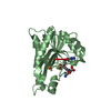
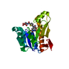
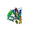
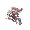
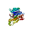
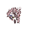
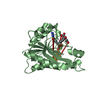
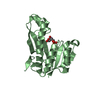
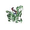
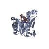
Human GAR transformylase in complex with GAR and (4-{[2-(2-Amino-4-oxo-4,7-dihydro-3H-pyrrolo[2,3-d]pyrimidin-6-yl)ethyl]amino}benzoyl)-L-glutamic acid (AGF183)
ComponentsTrifunctional purine biosynthetic protein adenosine-3
Keywordsligase/ligase inhibitor / gar transformylase / antifolate / ligase-ligase inhibitor complex
Function / homology
Function and homology information


phosphoribosylformylglycinamidine cyclo-ligase / phosphoribosylformylglycinamidine cyclo-ligase activity / adenine biosynthetic process / phosphoribosylamine-glycine ligase / phosphoribosylamine-glycine ligase activity / phosphoribosylglycinamide formyltransferase 1 / purine ribonucleoside monophosphate biosynthetic process / phosphoribosylglycinamide formyltransferase activity / 'de novo' XMP biosynthetic process / brainstem development ...phosphoribosylformylglycinamidine cyclo-ligase / phosphoribosylformylglycinamidine cyclo-ligase activity / adenine biosynthetic process / phosphoribosylamine-glycine ligase / phosphoribosylamine-glycine ligase activity / phosphoribosylglycinamide formyltransferase 1 / purine ribonucleoside monophosphate biosynthetic process / phosphoribosylglycinamide formyltransferase activity / 'de novo' XMP biosynthetic process / brainstem development / Purine ribonucleoside monophosphate biosynthesis / glycine metabolic process / 'de novo' AMP biosynthetic process / GMP biosynthetic process / purine nucleotide biosynthetic process / 'de novo' IMP biosynthetic process / : / tetrahydrofolate biosynthetic process / cerebellum development / : / cerebral cortex development / extracellular exosome / ATP binding / metal ion binding / cytosol
Similarity search - Function
Phosphoribosylformylglycinamidine cyclo-ligase / Phosphoribosylglycinamide synthetase, ATP-grasp (A) domain / Phosphoribosylglycinamide synthetase / Phosphoribosylglycinamide synthetase, conserved site / Phosphoribosylglycinamide synthetase, C-domain / Phosphoribosylglycinamide synthetase, N-terminal / Phosphoribosylglycinamide synthetase, C-domain superfamily / Phosphoribosylglycinamide synthetase, C domain / Phosphoribosylglycinamide synthetase, N domain / Phosphoribosylglycinamide synthetase signature. ...Phosphoribosylformylglycinamidine cyclo-ligase / Phosphoribosylglycinamide synthetase, ATP-grasp (A) domain / Phosphoribosylglycinamide synthetase / Phosphoribosylglycinamide synthetase, conserved site / Phosphoribosylglycinamide synthetase, C-domain / Phosphoribosylglycinamide synthetase, N-terminal / Phosphoribosylglycinamide synthetase, C-domain superfamily / Phosphoribosylglycinamide synthetase, C domain / Phosphoribosylglycinamide synthetase, N domain / Phosphoribosylglycinamide synthetase signature. / Phosphoribosylglycinamide synthetase, C domain / Phosphoribosylglycinamide synthetase, ATP-grasp (A) domain / Phosphoribosylglycinamide synthetase, ATP-grasp (A) domain / Phosphoribosylglycinamide formyltransferase / Formyl transferase, N-terminal domain / PurM-like, N-terminal domain / AIR synthase related protein, N-terminal domain / Phosphoribosylglycinamide formyltransferase, active site / Phosphoribosylglycinamide formyltransferase active site. / PurM-like, C-terminal domain / PurM-like, C-terminal domain superfamily / PurM-like, N-terminal domain superfamily / AIR synthase related protein, C-terminal domain / Formyl transferase, N-terminal / Formyl transferase / Formyl transferase, N-terminal domain superfamily / Rudiment single hybrid motif / ATP-grasp fold, subdomain 1 / Pre-ATP-grasp domain superfamily / ATP-grasp fold / ATP-grasp fold profile. / Rossmann fold / 3-Layer(aba) Sandwich / Alpha Beta
Similarity search - Domain/homology
Chem-83A / GLYCINAMIDE RIBONUCLEOTIDE / Trifunctional purine biosynthetic protein adenosine-3
Similarity search - Component
Biological speciesHomo sapiens (human)
MethodX-RAY DIFFRACTION / SYNCHROTRON / MOLECULAR REPLACEMENT / Resolution: 2.1 Å
AuthorsWong, J. / Deis, S.M. / Dann III, C.E.
Funding support United States, 2items
OrganizationGrant numberCountry
National Institutes of Health/National Cancer Institute (NIH/NCI)CA166711 United States
National Institutes of Health/National Institute of General Medical Sciences (NIH/NIGMS)GM094472 United States
CitationJournal: J.Med.Chem. / Year: 2016
Title: Tumor Targeting with Novel 6-Substituted Pyrrolo [2,3-d] Pyrimidine Antifolates with Heteroatom Bridge Substitutions via Cellular Uptake by Folate Receptor alpha and the Proton-Coupled Folate ...Title: Tumor Targeting with Novel 6-Substituted Pyrrolo [2,3-d] Pyrimidine Antifolates with Heteroatom Bridge Substitutions via Cellular Uptake by Folate Receptor alpha and the Proton-Coupled Folate Transporter and Inhibition of de Novo Purine Nucleotide Biosynthesis.
Authors: Golani, L.K. / Wallace-Povirk, A. / Deis, S.M. / Wong, J. / Ke, J. / Gu, X. / Raghavan, S. / Wilson, M.R. / Li, X. / Polin, L. / de Waal, P.W. / White, K. / Kushner, J. / O'Connor, C. / Hou, ...Authors: Golani, L.K. / Wallace-Povirk, A. / Deis, S.M. / Wong, J. / Ke, J. / Gu, X. / Raghavan, S. / Wilson, M.R. / Li, X. / Polin, L. / de Waal, P.W. / White, K. / Kushner, J. / O'Connor, C. / Hou, Z. / Xu, H.E. / Melcher, K. / Dann, C.E. / Matherly, L.H. / Gangjee, A.
History
DepositionApr 9, 2016Deposition site: RCSB / Processing site: RCSB
Revision 1.0Aug 10, 2016Provider: repository / Type: Initial release
Revision 1.1Sep 21, 2016Group: Database references
Revision 1.2Sep 20, 2017Group: Author supporting evidence / Database references / Derived calculations
Category: citation / pdbx_audit_support / pdbx_struct_oper_list
Item: _citation.journal_id_CSD / _pdbx_audit_support.funding_organization / _pdbx_struct_oper_list.symmetry_operation
Revision 1.3Dec 4, 2019Group: Author supporting evidence / Category: pdbx_audit_support / Item: _pdbx_audit_support.funding_organization
Revision 1.4Sep 27, 2023Group: Data collection / Database references / Refinement description
Category: chem_comp_atom / chem_comp_bond ...chem_comp_atom / chem_comp_bond / database_2 / diffrn_radiation_wavelength / pdbx_initial_refinement_model
Item: _database_2.pdbx_DOI / _database_2.pdbx_database_accession

-
Structure visualization

Structure viewerMolecule:
MolmilJmol/JSmol

Downloads & links

-
Assembly

Deposited unit
A: Trifunctional purine biosynthetic protein adenosine-3
hetero molecules


Theoretical massNumber of molelcules
Total (without water)23,4803
Polymers22,7531
Non-polymers7272
Water93752
1


  • Idetical with deposited unit
  • defined by author&software
TypeNameSymmetry operationNumber
identity operation1_555x,y,z1
MethodPISA
2


  • Idetical with deposited unit
  • defined by author&software
TypeNameSymmetry operationNumber
identity operation1_555x,y,z1
Buried area610 Å2
ΔGint-5 kcal/mol
Surface area9340 Å2
MethodPISA
Unit cell
Length a, b, c (Å)75.386, 75.386, 101.293
Angle α, β, γ (deg.)90.00, 90.00, 120.00
Int Tables number154
Space group name H-MP3221

-
Components

#1: Protein Trifunctional purine biosynthetic protein adenosine-3


Mass: 22753.088 Da / Num. of mol.: 1 / Fragment: unp residues 808-1010 / Mutation: Q905A
Source method: isolated from a genetically manipulated source
Source: (gene. exp.) Homo sapiens (human) / Gene: GART, PGFT, PRGS / Plasmid: pET22B / Production host: Escherichia coli (E. coli) / Strain (production host): Rosetta(DE3)pLysS
References: UniProt: P22102, phosphoribosylamine-glycine ligase, phosphoribosylformylglycinamidine cyclo-ligase, phosphoribosylglycinamide formyltransferase 1
#2: Chemical ChemComp-GAR / GLYCINAMIDE RIBONUCLEOTIDE


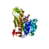
Mass: 284.160 Da / Num. of mol.: 1 / Source method: isolated from a natural source / Formula: C7H13N2O8P
#3: Chemical ChemComp-83A / N-(4-{[2-(2-amino-4-oxo-4,7-dihydro-3H-pyrrolo[2,3-d]pyrimidin-6-yl)ethyl]amino}benzene-1-carbonyl)-L-glutamic acid / antifolate AGF183


Mass: 442.425 Da / Num. of mol.: 1 / Source method: obtained synthetically / Formula: C20H22N6O6
#4: Water ChemComp-HOH / water


Mass: 18.015 Da / Num. of mol.: 52 / Source method: isolated from a natural source / Formula: H2O

-
Experimental details

-
Experiment

ExperimentMethod: X-RAY DIFFRACTION / Number of used crystals: 1

-
Sample preparation

CrystalDensity Matthews: 3.64 Å3/Da / Density % sol: 66.23 %
Crystal growTemperature: 277 K / Method: vapor diffusion, sitting drop / pH: 7.5
Details: 0.1 M Tris (pH 7.5), 0.333 mM NaCl, 18% polyethylene glycol (PEG) 3350, and 2% PEG 400

-
Data collection

DiffractionMean temperature: 110 K
Diffraction sourceSource: SYNCHROTRON / Site: APS / Beamline: 24-ID-E / Wavelength: 1 Å
DetectorType: ADSC QUANTUM 315 / Detector: CCD / Date: Dec 9, 2015
RadiationProtocol: SINGLE WAVELENGTH / Monochromatic (M) / Laue (L): M / Scattering type: x-ray
Radiation wavelengthWavelength: 1 Å / Relative weight: 1
ReflectionResolution: 2.1→40.017 Å / Num. obs: 22067 / % possible obs: 100 % / Redundancy: 5.4 % / Rmerge(I) obs: 0.1 / Net I/σ(I): 13
Reflection shellResolution: 2.1→2.16 Å / Redundancy: 5.5 % / Rmerge(I) obs: 0.75 / Mean I/σ(I) obs: 2.6 / % possible all: 100

-
Processing

Software
NameVersionClassification
PHENIX1.9_1692refinement
XDSdata reduction
XDSdata scaling
PHASERphasing
RefinementMethod to determine structure: MOLECULAR REPLACEMENT
Starting model: 4ZZ1
Resolution: 2.1→40.02 Å / SU ML: 0.22 / Cross valid method: FREE R-VALUE / σ(F): 1.35 / Phase error: 20.41 / Stereochemistry target values: ML
RfactorNum. reflection% reflection
Rfree0.213 1825 9.15 %
Rwork0.174 --
obs0.177 19940 99.9 %
Solvent computationShrinkage radii: 0.9 Å / VDW probe radii: 1.11 Å / Solvent model: FLAT BULK SOLVENT MODEL
Refinement stepCycle: LAST / Resolution: 2.1→40.02 Å
ProteinNucleic acidLigandSolventTotal
Num. atoms1499 0 50 52 1601
Refine LS restraints
Refine-IDTypeDev idealNumber
X-RAY DIFFRACTIONf_bond_d0.0141574
X-RAY DIFFRACTIONf_angle_d1.4762139
X-RAY DIFFRACTIONf_dihedral_angle_d13.722564
X-RAY DIFFRACTIONf_chiral_restr0.072256
X-RAY DIFFRACTIONf_plane_restr0.006271
LS refinement shell
Resolution (Å)Rfactor RfreeNum. reflection RfreeRfactor RworkNum. reflection RworkRefine-ID% reflection obs (%)
2.1-2.15680.27231390.22591361X-RAY DIFFRACTION100
2.1568-2.22030.26371390.19661374X-RAY DIFFRACTION100
2.2203-2.29190.22331350.1911373X-RAY DIFFRACTION100
2.2919-2.37380.22581380.17611377X-RAY DIFFRACTION100
2.3738-2.46890.21491380.18461371X-RAY DIFFRACTION100
2.4689-2.58120.21841350.18161381X-RAY DIFFRACTION100
2.5812-2.71730.26521350.19451384X-RAY DIFFRACTION100
2.7173-2.88750.25681410.20811384X-RAY DIFFRACTION100
2.8875-3.11030.27011420.20631400X-RAY DIFFRACTION100
3.1103-3.42320.23261480.20091395X-RAY DIFFRACTION100
3.4232-3.91820.19431420.16731396X-RAY DIFFRACTION100
3.9182-4.9350.15761420.12851419X-RAY DIFFRACTION100
4.935-40.02420.17711510.14261500X-RAY DIFFRACTION99
Refinement TLS params.Method: refined / Origin x: 31.716 Å / Origin y: -19.1007 Å / Origin z: -22.6729 Å
111213212223313233
T0.173 Å2-0.0108 Å20.0318 Å2-0.1388 Å20.0117 Å2--0.1817 Å2
L1.0309 °20.1305 °2-0.6686 °2-0.8954 °2-0.23 °2--1.4334 °2
S0.1441 Å °0.0173 Å °0.2381 Å °0.0145 Å °-0.0167 Å °-0.0368 Å °-0.1956 Å °0.0272 Å °0.0058 Å °
Refinement TLS groupSelection details: ALL

+
About Yorodumi

-
News

-
Feb 9, 2022. New format data for meta-information of EMDB entries

New format data for meta-information of EMDB entries

  • Version 3 of the EMDB header file is now the official format.
  • The previous official version 1.9 will be removed from the archive.

Related info.:EMDB header

External links:wwPDB to switch to version 3 of the EMDB data model

-
Aug 12, 2020. Covid-19 info

Covid-19 info

URL: https://pdbjlvh1.pdbj.org/emnavi/covid19.php

New page: Covid-19 featured information page in EM Navigator.

Related info.:Covid-19 info / Mar 5, 2020. Novel coronavirus structure data

+
Mar 5, 2020. Novel coronavirus structure data

Novel coronavirus structure data

Related info.:Yorodumi Speices / Aug 12, 2020. Covid-19 info

External links:COVID-19 featured content - PDBj / Molecule of the Month (242):Coronavirus Proteases

+
Jan 31, 2019. EMDB accession codes are about to change! (news from PDBe EMDB page)

EMDB accession codes are about to change! (news from PDBe EMDB page)

  • The allocation of 4 digits for EMDB accession codes will soon come to an end. Whilst these codes will remain in use, new EMDB accession codes will include an additional digit and will expand incrementally as the available range of codes is exhausted. The current 4-digit format prefixed with “EMD-” (i.e. EMD-XXXX) will advance to a 5-digit format (i.e. EMD-XXXXX), and so on. It is currently estimated that the 4-digit codes will be depleted around Spring 2019, at which point the 5-digit format will come into force.
  • The EM Navigator/Yorodumi systems omit the EMD- prefix.

Related info.:Q: What is EMD? / ID/Accession-code notation in Yorodumi/EM Navigator

External links:EMDB Accession Codes are Changing Soon! / Contact to PDBj

+
Jul 12, 2017. Major update of PDB

Major update of PDB

  • wwPDB released updated PDB data conforming to the new PDBx/mmCIF dictionary.
  • This is a major update changing the version number from 4 to 5, and with Remediation, in which all the entries are updated.
  • In this update, many items about electron microscopy experimental information are reorganized (e.g. em_software).
  • Now, EM Navigator and Yorodumi are based on the updated data.

External links:wwPDB Remediation / Enriched Model Files Conforming to OneDep Data Standards Now Available in the PDB FTP Archive

-
Yorodumi

Thousand views of thousand structures

  • Yorodumi is a browser for structure data from EMDB, PDB, SASBDB, etc.
  • This page is also the successor to EM Navigator detail page, and also detail information page/front-end page for Omokage search.
  • The word "yorodu" (or yorozu) is an old Japanese word meaning "ten thousand". "mi" (miru) is to see.

Related info.:EMDB / PDB / SASBDB / Comparison of 3 databanks / Yorodumi Search / Aug 31, 2016. New EM Navigator & Yorodumi / Yorodumi Papers / Jmol/JSmol / Function and homology information / Changes in new EM Navigator and Yorodumi

Read more