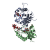
 Movie
Movie Controller


+ Open data

- Basic information
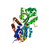
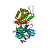
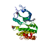
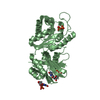
| Entry | Database: PDB / ID: 1r9q | ||||||
|---|---|---|---|---|---|---|---|
| Title | structure analysis of ProX in complex with proline betaine | ||||||
 Components Components | Glycine betaine-binding periplasmic protein | ||||||
 Keywords Keywords | PROTEIN BINDING / periplasmic binding protein / cation-pi interactions / tryptophan box | ||||||
| Function / homology |  Function and homology information Function and homology informationProVWX complex / quaternary ammonium group binding / glycine betaine transport / glycine import across plasma membrane / amino acid import across plasma membrane / cellular response to osmotic stress / hyperosmotic response / amino acid transport / transmembrane transporter activity / ATP-binding cassette (ABC) transporter complex ...ProVWX complex / quaternary ammonium group binding / glycine betaine transport / glycine import across plasma membrane / amino acid import across plasma membrane / cellular response to osmotic stress / hyperosmotic response / amino acid transport / transmembrane transporter activity / ATP-binding cassette (ABC) transporter complex / outer membrane-bounded periplasmic space / periplasmic space / membrane Similarity search - Function | ||||||
| Biological species |  | ||||||
| Method |  X-RAY DIFFRACTION / MOLECULAR REPLACEMENT / Resolution: 2.05 Å X-RAY DIFFRACTION / MOLECULAR REPLACEMENT / Resolution: 2.05 Å | ||||||
 Authors Authors | Schiefner, A. / Breed, J. / Bosser, L. / Kneip, S. / Gade, J. / Holtmann, G. / Diederichs, K. / Welte, W. / Bremer, E. | ||||||
 Citation Citation | Journal: J.BIOL.CHEM. / Year: 2004 Title: Cation-pi Interactions as Determinants for Binding of the Compatible Solutes Glycine Betaine and Proline Betaine by the Periplasmic Ligand-binding Protein ProX from Escherichia coli Authors: Schiefner, A. / Breed, J. / Bosser, L. / Kneip, S. / Gade, J. / Holtmann, G. / Diederichs, K. / Welte, W. / Bremer, E. | ||||||
| History |
|
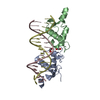
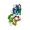
- Structure visualization
| Structure viewer | Molecule: MolmilJmol/JSmol |
|---|
- Downloads & links
-Download
| PDBx/mmCIF format | 1r9q.cif.gz | 72.6 KB | Display | PDBx/mmCIF format |
|---|---|---|---|---|
| PDB format | pdb1r9q.ent.gz | 53.9 KB | Display | PDB format |
| PDBx/mmJSON format | 1r9q.json.gz | Tree view | PDBx/mmJSON format | |
| Others |  Other downloads Other downloads |
-Validation report
| Arichive directory | https://data.pdbj.org/pub/pdb/validation_reports/r9/1r9qftp://data.pdbj.org/pub/pdb/validation_reports/r9/1r9q | HTTPS FTP |
|---|
-Related structure data
| Related structure data |  1r9lSC S: Starting model for refinement C: citing same article ( |
|---|---|
| Similar structure data |


-Links
 PDBj
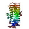
PDBj- Assembly
| Deposited unit | 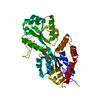
| ||||||||
|---|---|---|---|---|---|---|---|---|---|
| 1 |
| ||||||||
| Unit cell |
|
-Components
| #1: Protein | Mass: 33760.742 Da / Num. of mol.: 1 Source method: isolated from a genetically manipulated source Source: (gene. exp.) |
|---|---|
| #2: Chemical | ChemComp-UNX /   Num. of mol.: 1 / Source method: obtained synthetically Num. of mol.: 1 / Source method: obtained synthetically |
| #3: Chemical | ChemComp-PBE / 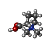  Mass: 144.192 Da / Num. of mol.: 1 / Source method: obtained synthetically / Formula: C7H14NO2 Mass: 144.192 Da / Num. of mol.: 1 / Source method: obtained synthetically / Formula: C7H14NO2 |
| #4: Water | ChemComp-HOH /  Mass: 18.015 Da / Num. of mol.: 59 / Source method: isolated from a natural source / Formula: H2O Mass: 18.015 Da / Num. of mol.: 59 / Source method: isolated from a natural source / Formula: H2O |
-Experimental details
-Experiment
| Experiment | Method: X-RAY DIFFRACTION / Number of used crystals: 1 |
|---|
- Sample preparation
| Crystal | Density Matthews: 2.21 Å3/Da / Density % sol: 43.96 % | ||||||||||||||||||||||||||||||
|---|---|---|---|---|---|---|---|---|---|---|---|---|---|---|---|---|---|---|---|---|---|---|---|---|---|---|---|---|---|---|---|
| Crystal grow | Temperature: 291 K / Method: vapor diffusion, hanging drop / pH: 6.3 Details: PEG 4000, PIPES, pH 6.3, VAPOR DIFFUSION, HANGING DROP, temperature 291K | ||||||||||||||||||||||||||||||
| Crystal grow | *PLUS Temperature: 18 ℃ / pH: 8.3 / Method: vapor diffusion | ||||||||||||||||||||||||||||||
| Components of the solutions | *PLUS
|
-Data collection
| Diffraction | Mean temperature: 291 K |
|---|---|
| Diffraction source | Source: ROTATING ANODE / Wavelength: 1.54179 Å |
| Detector | Type: MARRESEARCH / Detector: IMAGE PLATE / Date: Jan 21, 2000 |
| Radiation | Protocol: SINGLE WAVELENGTH / Monochromatic (M) / Laue (L): M / Scattering type: x-ray |
| Radiation wavelength | Wavelength: 1.54179 Å / Relative weight: 1 |
| Reflection | Resolution: 2.05→15 Å / Num. all: 19587 / Num. obs: 19587 / % possible obs: 99 % / Observed criterion σ(F): 0 / Observed criterion σ(I): 0 |
| Reflection shell | Resolution: 2.05→2.25 Å / % possible all: 99.4 |
| Reflection | *PLUS Lowest resolution: 15 Å / Num. measured all: 75377 / Rmerge(I) obs: 0.06 |
| Reflection shell | *PLUS % possible obs: 99.4 % / Rmerge(I) obs: 0.239 / Mean I/σ(I) obs: 5.65 |
- Processing
| Software |
| ||||||||||||||||||||
|---|---|---|---|---|---|---|---|---|---|---|---|---|---|---|---|---|---|---|---|---|---|
| Refinement | Method to determine structure: MOLECULAR REPLACEMENT Starting model: 1R9L Resolution: 2.05→10 Å / σ(F): 0 / σ(I): 0 / Stereochemistry target values: Engh & Huber
| ||||||||||||||||||||
| Refinement step | Cycle: LAST / Resolution: 2.05→10 Å
| ||||||||||||||||||||
| LS refinement shell | Resolution: 2.08→2.13 Å / Rfactor Rfree error: 0
| ||||||||||||||||||||
| Refinement | *PLUS Lowest resolution: 15 Å | ||||||||||||||||||||
| Solvent computation | *PLUS | ||||||||||||||||||||
| Displacement parameters | *PLUS | ||||||||||||||||||||
| Refine LS restraints | *PLUS
|
