 Movie
Movie Controller


+ Open data

- Basic information
| Entry | Database: PDB / ID: 1r7a | ||||||
|---|---|---|---|---|---|---|---|
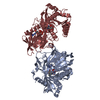
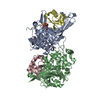
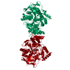
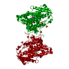
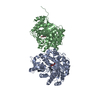
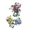
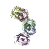
| Title | Sucrose Phosphorylase from Bifidobacterium adolescentis | ||||||
 Components Components | sucrose phosphorylase | ||||||
 Keywords Keywords | TRANSFERASE / beta-alpha-barrels / dimer / glycoside hydrolase | ||||||
| Function / homology |  Function and homology information Function and homology informationsucrose phosphorylase / sucrose phosphorylase activity / carbohydrate metabolic process Similarity search - Function | ||||||
| Biological species |  Bifidobacterium adolescentis (bacteria) Bifidobacterium adolescentis (bacteria) | ||||||
| Method |  X-RAY DIFFRACTION / SYNCHROTRON / SIRAS / Resolution: 1.77 Å X-RAY DIFFRACTION / SYNCHROTRON / SIRAS / Resolution: 1.77 Å | ||||||
 Authors Authors | Sprogoe, D. / van den Broek, L.A.M. / Mirza, O. / Kastrup, J.S. / Voragen, A.G.J. / Gajhede, M. / Skov, L.K. | ||||||
 Citation Citation | Journal: Biochemistry / Year: 2004 Title: Crystal structure of sucrose phosphorylase from Bifidobacterium adolescentis. Authors: Sprogoe, D. / van den Broek, L.A. / Mirza, O. / Kastrup, J.S. / Voragen, A.G. / Gajhede, M. / Skov, L.K. | ||||||
| History |
|
- Structure visualization
| Structure viewer | Molecule: MolmilJmol/JSmol |
|---|
- Downloads & links
-Download
| PDBx/mmCIF format | 1r7a.cif.gz | 232.2 KB | Display | PDBx/mmCIF format |
|---|---|---|---|---|
| PDB format | pdb1r7a.ent.gz | 193.7 KB | Display | PDB format |
| PDBx/mmJSON format | 1r7a.json.gz | Tree view | PDBx/mmJSON format | |
| Others |  Other downloads Other downloads |
-Validation report
| Summary document | 1r7a_validation.pdf.gz | 452.9 KB | Display | wwPDB validaton report |
|---|---|---|---|---|
| Full document | 1r7a_full_validation.pdf.gz | 459.6 KB | Display | |
| Data in XML | 1r7a_validation.xml.gz | 50.4 KB | Display | |
| Data in CIF | 1r7a_validation.cif.gz | 78.2 KB | Display | |
| Arichive directory | https://data.pdbj.org/pub/pdb/validation_reports/r7/1r7aftp://data.pdbj.org/pub/pdb/validation_reports/r7/1r7a | HTTPS FTP |
-Related structure data
| Similar structure data |
|---|

-Links
 PDBj
PDBj

- Assembly
| Deposited unit | 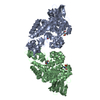
| ||||||||
|---|---|---|---|---|---|---|---|---|---|
| 1 |
| ||||||||
| Unit cell |
| ||||||||
| Details | The biological unit is probably a dimer (chains A and B) as given in the coordinates. |
-Components
| #1: Protein | Mass: 56284.668 Da / Num. of mol.: 2 Source method: isolated from a genetically manipulated source Source: (gene. exp.) Bifidobacterium adolescentis (bacteria)Plasmid: pBluescript / Production host: References: UniProt: Q84HQ2, UniProt: A0ZZH6*PLUS, sucrose phosphorylase #2: Chemical |   Mass: 122.143 Da / Num. of mol.: 2 / Source method: obtained synthetically / Formula: C4H12NO3 / Comment: pH buffer*YM Mass: 122.143 Da / Num. of mol.: 2 / Source method: obtained synthetically / Formula: C4H12NO3 / Comment: pH buffer*YM#3: Water | ChemComp-HOH / |  Mass: 18.015 Da / Num. of mol.: 1421 / Source method: isolated from a natural source / Formula: H2O Mass: 18.015 Da / Num. of mol.: 1421 / Source method: isolated from a natural source / Formula: H2O |
|---|
-Experimental details
-Experiment
| Experiment | Method: X-RAY DIFFRACTION / Number of used crystals: 3 |
|---|
- Sample preparation
| Crystal | Density Matthews: 2.28 Å3/Da / Density % sol: 46.13 % | ||||||||||||||||||||||||||||||||||||
|---|---|---|---|---|---|---|---|---|---|---|---|---|---|---|---|---|---|---|---|---|---|---|---|---|---|---|---|---|---|---|---|---|---|---|---|---|---|
| Crystal grow | Temperature: 298 K / Method: vapor diffusion, hanging drop / pH: 8 Details: PEG 4000, sodium acetate, tris, pH 8, VAPOR DIFFUSION, HANGING DROP, temperature 298K | ||||||||||||||||||||||||||||||||||||
| Crystal grow | *PLUS Temperature: 25 ℃ / pH: 7.1 / Method: vapor diffusion, hanging drop | ||||||||||||||||||||||||||||||||||||
| Components of the solutions | *PLUS
|
-Data collection
| Diffraction |
| ||||||||||||||||||||||||
|---|---|---|---|---|---|---|---|---|---|---|---|---|---|---|---|---|---|---|---|---|---|---|---|---|---|
| Diffraction source |
| ||||||||||||||||||||||||
| Detector |
| ||||||||||||||||||||||||
| Radiation |
| ||||||||||||||||||||||||
| Radiation wavelength |
| ||||||||||||||||||||||||
| Reflection | Resolution: 1.58→23.3 Å / Num. all: 138899 / Num. obs: 138899 / % possible obs: 97.8 % / Observed criterion σ(F): 0 / Observed criterion σ(I): 0 / Redundancy: 3.7 % / Biso Wilson estimate: 10.4 Å2 / Rsym value: 0.109 / Net I/σ(I): 4.3 | ||||||||||||||||||||||||
| Reflection shell | Resolution: 1.58→1.66 Å / Redundancy: 3.2 % / Mean I/σ(I) obs: 0.9 / Num. unique all: 17475 / Rsym value: 0.53 / % possible all: 97.8 | ||||||||||||||||||||||||
| Reflection | *PLUS Rmerge(I) obs: 0.109 | ||||||||||||||||||||||||
| Reflection shell | *PLUS % possible obs: 85.2 % / Rmerge(I) obs: 0.53 |


- Processing
| Software |
| ||||||||||||||||||||||||||||||||||||
|---|---|---|---|---|---|---|---|---|---|---|---|---|---|---|---|---|---|---|---|---|---|---|---|---|---|---|---|---|---|---|---|---|---|---|---|---|---|
| Refinement | Method to determine structure: SIRAS / Resolution: 1.77→19.88 Å / Rfactor Rfree error: 0.003 / Data cutoff high absF: 2663906.59 / Data cutoff low absF: 0 / Isotropic thermal model: RESTRAINED / Cross valid method: THROUGHOUT / σ(F): 0 / Stereochemistry target values: Engh & Huber
| ||||||||||||||||||||||||||||||||||||
| Solvent computation | Solvent model: FLAT MODEL / Bsol: 50.2152 Å2 / ksol: 0.379705 e/Å3 | ||||||||||||||||||||||||||||||||||||
| Displacement parameters | Biso mean: 14.8 Å2
| ||||||||||||||||||||||||||||||||||||
| Refine analyze |
| ||||||||||||||||||||||||||||||||||||
| Refinement step | Cycle: LAST / Resolution: 1.77→19.88 Å
| ||||||||||||||||||||||||||||||||||||
| Refine LS restraints |
| ||||||||||||||||||||||||||||||||||||
| LS refinement shell | Resolution: 1.77→1.88 Å / Rfactor Rfree error: 0.007 / Total num. of bins used: 6
| ||||||||||||||||||||||||||||||||||||
| Xplor file |
| ||||||||||||||||||||||||||||||||||||
| Refinement | *PLUS Lowest resolution: 20 Å / Num. reflection obs: 101196 / % reflection Rfree: 5 % / Rfactor Rfree: 0.191 / Rfactor Rwork: 0.158 | ||||||||||||||||||||||||||||||||||||
| Solvent computation | *PLUS | ||||||||||||||||||||||||||||||||||||
| Displacement parameters | *PLUS | ||||||||||||||||||||||||||||||||||||
| Refine LS restraints | *PLUS
|
