 Movie
Movie Controller

[English] 日本語
 Yorodumi
- PDB-1r6f: The structure of Yersinia pestis V-antigen, an essential virulenc... -
+ Open data

- Basic information
| Entry | Database: PDB / ID: 1r6f | ||||||
|---|---|---|---|---|---|---|---|
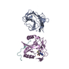
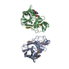
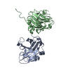
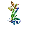
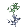
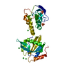
| Title | The structure of Yersinia pestis V-antigen, an essential virulence factor and mediator of immunity against plague | ||||||
 Components Components | Virulence-associated V antigen | ||||||
 Keywords Keywords | PROTEIN BINDING / coiled-coil | ||||||
| Function / homology | Low calcium response V antigen / Virulence-associated V antigen superfamily / V antigen (LcrV) protein / : / extracellular region / Virulence-associated V antigen / Virulence-associated V antigen Function and homology information Function and homology information | ||||||
| Biological species |   Yersinia pestis (bacteria) Yersinia pestis (bacteria) | ||||||
| Method |  X-RAY DIFFRACTION / SYNCHROTRON / MAD / Resolution: 2.17 Å X-RAY DIFFRACTION / SYNCHROTRON / MAD / Resolution: 2.17 Å | ||||||
 Authors Authors | Derewenda, U. / Mateja, A. / Devedjiev, Y. / Routzahn, K.M. / Evdokimov, A.G. / Derewenda, Z.S. / Waugh, D.S. | ||||||
 Citation Citation | Journal: Structure / Year: 2004 Title: The structure of Yersinia pestis V-antigen, an essential virulence factor and mediator of immunity against plague Authors: Derewenda, U. / Mateja, A. / Devedjiev, Y. / Routzahn, K.M. / Evdokimov, A.G. / Derewenda, Z.S. / Waugh, D.S. | ||||||
| History |
|
- Structure visualization
| Structure viewer | Molecule: MolmilJmol/JSmol |
|---|
- Downloads & links
-Download
| PDBx/mmCIF format | 1r6f.cif.gz | 64.2 KB | Display | PDBx/mmCIF format |
|---|---|---|---|---|
| PDB format | pdb1r6f.ent.gz | 46.9 KB | Display | PDB format |
| PDBx/mmJSON format | 1r6f.json.gz | Tree view | PDBx/mmJSON format | |
| Others |  Other downloads Other downloads |
-Validation report
| Arichive directory | https://data.pdbj.org/pub/pdb/validation_reports/r6/1r6fftp://data.pdbj.org/pub/pdb/validation_reports/r6/1r6f | HTTPS FTP |
|---|
-Related structure data
| Similar structure data |
|---|


-Links
 PDBj
PDBj- Assembly
| Deposited unit | 
| ||||||||
|---|---|---|---|---|---|---|---|---|---|
| 1 |
| ||||||||
| Unit cell |
|
-Components
| #1: Protein | Mass: 35158.555 Da / Num. of mol.: 1 / Fragment: Lcrv fragment 24-323 / Mutation: K40A D41A K42A C273S Source method: isolated from a genetically manipulated source Source: (gene. exp.) Yersinia pestis (bacteria) / Plasmid: MBP-LCRV(DELTA1-23-C273S)-HIS6 / Production host: |
|---|
-Experimental details
-Experiment
| Experiment | Method: X-RAY DIFFRACTION |
|---|
- Sample preparation
| Crystal | Density Matthews: 2.02 Å3/Da / Density % sol: 39.07 % | ||||||||||||||||||||||||||||||
|---|---|---|---|---|---|---|---|---|---|---|---|---|---|---|---|---|---|---|---|---|---|---|---|---|---|---|---|---|---|---|---|
| Crystal grow | Temperature: 294 K / Method: vapor diffusion, hanging drop / pH: 8.7 Details: PEG 5000 MME, sodium acetate, Tris, pH 8.7, VAPOR DIFFUSION, HANGING DROP, temperature 294K | ||||||||||||||||||||||||||||||
| Crystal grow | *PLUS Method: vapor diffusion, sitting drop | ||||||||||||||||||||||||||||||
| Components of the solutions | *PLUS
|
-Data collection
| Diffraction |
| |||||||||||||||
|---|---|---|---|---|---|---|---|---|---|---|---|---|---|---|---|---|
| Diffraction source |
| |||||||||||||||
| Detector |
| |||||||||||||||
| Radiation | Monochromator: Si(111) / Protocol: MAD / Monochromatic (M) / Laue (L): M / Scattering type: x-ray | |||||||||||||||
| Radiation wavelength |
| |||||||||||||||
| Reflection | Resolution: 2.17→18.5 Å / Num. all: 14116 / Num. obs: 14116 / % possible obs: 95.9 % / Observed criterion σ(F): -3 / Observed criterion σ(I): -3 / Redundancy: 2.2 % / Rmerge(I) obs: 0.077 / Rsym value: 0.077 / Net I/σ(I): 12.5 | |||||||||||||||
| Reflection shell | Resolution: 2.17→2.25 Å / Rmerge(I) obs: 0.37 / Mean I/σ(I) obs: 2 / % possible all: 86.2 | |||||||||||||||
| Reflection | *PLUS Num. measured all: 31607 | |||||||||||||||
| Reflection shell | *PLUS % possible obs: 86.2 % / Rmerge(I) obs: 0.37 |

- Processing
| Software |
| ||||||||||||||||||||||||||||||||||||||||||||||||||||||||||||||||||||||
|---|---|---|---|---|---|---|---|---|---|---|---|---|---|---|---|---|---|---|---|---|---|---|---|---|---|---|---|---|---|---|---|---|---|---|---|---|---|---|---|---|---|---|---|---|---|---|---|---|---|---|---|---|---|---|---|---|---|---|---|---|---|---|---|---|---|---|---|---|---|---|---|
| Refinement | Method to determine structure: MAD / Resolution: 2.17→18.5 Å / Cor.coef. Fo:Fc: 0.941 / Isotropic thermal model: Isotropic / σ(F): 0 / ESU R: 0.398
| ||||||||||||||||||||||||||||||||||||||||||||||||||||||||||||||||||||||
| Solvent computation | Ion probe radii: 0.8 Å / Shrinkage radii: 0.8 Å / VDW probe radii: 1.4 Å / Solvent model: BABINET MODEL WITH MASK | ||||||||||||||||||||||||||||||||||||||||||||||||||||||||||||||||||||||
| Displacement parameters | Biso mean: 27.251 Å2
| ||||||||||||||||||||||||||||||||||||||||||||||||||||||||||||||||||||||
| Refinement step | Cycle: LAST / Resolution: 2.17→18.5 Å
| ||||||||||||||||||||||||||||||||||||||||||||||||||||||||||||||||||||||
| Refine LS restraints |
| ||||||||||||||||||||||||||||||||||||||||||||||||||||||||||||||||||||||
| LS refinement shell | Resolution: 2.17→2.207 Å / Total num. of bins used: 20 /
| ||||||||||||||||||||||||||||||||||||||||||||||||||||||||||||||||||||||
| Refinement | *PLUS Lowest resolution: 30 Å | ||||||||||||||||||||||||||||||||||||||||||||||||||||||||||||||||||||||
| Solvent computation | *PLUS | ||||||||||||||||||||||||||||||||||||||||||||||||||||||||||||||||||||||
| Displacement parameters | *PLUS | ||||||||||||||||||||||||||||||||||||||||||||||||||||||||||||||||||||||
| Refine LS restraints | *PLUS
|
