 Movie
Movie Controller

[English] 日本語
 Yorodumi
- PDB-1r68: Role of the amino sugar in DNA binding of disaccharide anthracycl... -
+ Open data

- Basic information
| Entry | Database: PDB / ID: 1r68 | ||||||||||||||||||
|---|---|---|---|---|---|---|---|---|---|---|---|---|---|---|---|---|---|---|---|
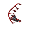
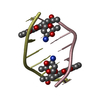
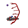
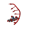
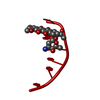
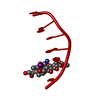
| Title | Role of the amino sugar in DNA binding of disaccharide anthracyclines: crystal structure of MAR70/d(CGATCG) complex | ||||||||||||||||||
 Components Components | 5'-D(* Keywords KeywordsDNA / right handed DNA / double helix / drug-DNA complex | Function / homology | 4'-EPI-4'-(2-DEOXYFUCOSE)DAUNOMYCIN / DNA |  Function and homology information Function and homology informationMethod |  X-RAY DIFFRACTION / MOLECULAR REPLACEMENT / Resolution: 1.2 Å X-RAY DIFFRACTION / MOLECULAR REPLACEMENT / Resolution: 1.2 Å  Authors AuthorsTemperini, C. / Cirilli, M. / Aschi, M. / Ughetto, G. |  CitationJournal: BIOORG.MED.CHEM. / Year: 2005 CitationJournal: BIOORG.MED.CHEM. / Year: 2005Title: Role of the amino sugar in DNA binding of disaccharide anthracyclines: crystal structure of the complex MAR70/d(CGATCG). Authors: Temperini, C. / Cirilli, M. / Aschi, M. / Ughetto, G. #1: Journal: Nucleic Acids Res. / Year: 2003Title: The crystal structure of the complex between a disaccharide anthracycline and the DNA hexamer d(CGATCG) reveals two different binding sites involving two DNA duplexes Authors: Temperini, C. / Messori, L. / Orioli, P. / Di Bugno, C. / Animati, F. / Ughetto, G. History |
|
- Structure visualization
| Structure viewer | Molecule: MolmilJmol/JSmol |
|---|
- Downloads & links
-Download
| PDBx/mmCIF format | 1r68.cif.gz | 17.2 KB | Display | PDBx/mmCIF format |
|---|---|---|---|---|
| PDB format | pdb1r68.ent.gz | 9.1 KB | Display | PDB format |
| PDBx/mmJSON format | 1r68.json.gz | Tree view | PDBx/mmJSON format | |
| Others |  Other downloads Other downloads |
-Validation report
| Arichive directory | https://data.pdbj.org/pub/pdb/validation_reports/r6/1r68ftp://data.pdbj.org/pub/pdb/validation_reports/r6/1r68 | HTTPS FTP |
|---|
-Related structure data
| Similar structure data |
|---|


-Links
 PDBj
PDBj

- Assembly
| Deposited unit | 
| ||||||||
|---|---|---|---|---|---|---|---|---|---|
| 1 | 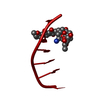
| ||||||||
| Unit cell |
|
-Components
| #1: DNA chain | Mass: 1809.217 Da / Num. of mol.: 1 / Source method: obtained synthetically |
|---|---|
| #2: Chemical | ChemComp-MAR /   Mass: 657.662 Da / Num. of mol.: 1 / Source method: obtained synthetically / Formula: C33H39NO13 Mass: 657.662 Da / Num. of mol.: 1 / Source method: obtained synthetically / Formula: C33H39NO13 |
| #3: Water | ChemComp-HOH /  Mass: 18.015 Da / Num. of mol.: 21 / Source method: isolated from a natural source / Formula: H2O Mass: 18.015 Da / Num. of mol.: 21 / Source method: isolated from a natural source / Formula: H2O |
-Experimental details
-Experiment
| Experiment | Method: X-RAY DIFFRACTION / Number of used crystals: 1 |
|---|
- Sample preparation
| Crystal | Density Matthews: 2.92 Å3/Da / Density % sol: 57.93 % | ||||||||||||||||||||||||||||||||||||||||||||
|---|---|---|---|---|---|---|---|---|---|---|---|---|---|---|---|---|---|---|---|---|---|---|---|---|---|---|---|---|---|---|---|---|---|---|---|---|---|---|---|---|---|---|---|---|---|
| Crystal grow | Temperature: 298 K / pH: 6 Details: Magnesium chloride, cacodylate, spermine, MPD, pH 6.0, VAPOR DIFFUSION, SITTING DROP, temperature 298K, pH 6.00 | ||||||||||||||||||||||||||||||||||||||||||||
| Components of the solutions |
|
-Data collection
| Diffraction | Mean temperature: 298 K |
|---|---|
| Diffraction source | Source: ROTATING ANODE / Type: RIGAKU RU200 / Wavelength: 1.54 |
| Detector | Type: RIGAKU AFC-5R / Detector: DIFFRACTOMETER / Date: Feb 15, 2003 |
| Radiation | Monochromator: GRAPHITE / Protocol: SINGLE WAVELENGTH / Monochromatic (M) / Laue (L): M / Scattering type: x-ray |
| Radiation wavelength | Wavelength: 1.54 Å / Relative weight: 1 |
| Reflection | Resolution: 1.2→15 Å / Num. obs: 6369 / % possible obs: 97.8 % / Biso Wilson estimate: 15.34 Å2 / Rmerge(I) obs: 0.114 / Net I/σ(I): 4.3 |
| Reflection shell | Resolution: 1.2→1.3 Å / % possible all: 87 |
- Processing
| Software |
| |||||||||||||||||||||||||||||||||
|---|---|---|---|---|---|---|---|---|---|---|---|---|---|---|---|---|---|---|---|---|---|---|---|---|---|---|---|---|---|---|---|---|---|---|
| Refinement | Method to determine structure: MOLECULAR REPLACEMENT / Resolution: 1.2→10 Å / σ(F): 4 /
| |||||||||||||||||||||||||||||||||
| Refinement step | Cycle: LAST / Resolution: 1.2→10 Å
| |||||||||||||||||||||||||||||||||
| Refine LS restraints |
|
