 Movie
Movie Controller

[English] 日本語
 Yorodumi
- PDB-1r5t: The Crystal Structure of Cytidine Deaminase CDD1, an Orphan C to ... -
+ Open data

- Basic information
| Entry | Database: PDB / ID: 1r5t | ||||||
|---|---|---|---|---|---|---|---|
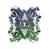
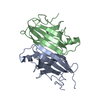
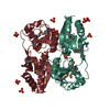
| Title | The Crystal Structure of Cytidine Deaminase CDD1, an Orphan C to U editase from Yeast | ||||||
 Components Components | Cytidine deaminase | ||||||
 Keywords Keywords | HYDROLASE / Zinc Dependent Deaminase / RNA editing / APOBEC-1 related protein | ||||||
| Function / homology |  Function and homology information Function and homology informationPyrimidine salvage / cytidine catabolic process / deoxycytidine catabolic process / pyrimidine-containing compound salvage / cytidine deaminase / cytidine deaminase activity / Neutrophil degranulation / zinc ion binding / identical protein binding / nucleus ...Pyrimidine salvage / cytidine catabolic process / deoxycytidine catabolic process / pyrimidine-containing compound salvage / cytidine deaminase / cytidine deaminase activity / Neutrophil degranulation / zinc ion binding / identical protein binding / nucleus / cytoplasm / cytosol Similarity search - Function | ||||||
| Biological species |  | ||||||
| Method |  X-RAY DIFFRACTION / SYNCHROTRON / MAD / Resolution: 2 Å X-RAY DIFFRACTION / SYNCHROTRON / MAD / Resolution: 2 Å | ||||||
 Authors Authors | Xie, K. / Sowden, M.P. / Dance, G.S.C. / Torelli, A.T. / Smith, H.C. / Wedekind, J.E. | ||||||
 Citation Citation | Journal: Proc.Natl.Acad.Sci.Usa / Year: 2004 Title: The structure of a yeast RNA-editing deaminase provides insight into the fold and function of activation-induced deaminase and APOBEC-1. Authors: Xie, K. / Sowden, M.P. / Dance, G.S. / Torelli, A.T. / Smith, H.C. / Wedekind, J.E. #1: Journal: Trends Genet. / Year: 2004Title: Activation induced deaminase: the importance of being specific Authors: Smith, H.C. / Bottaro, A. / Sowden, M.P. / Wedekind, J.E. #2: Journal: Trends Genet. / Year: 2003Title: Messenger RNA editing in mammals: new members of the APOBEC family seeking roles in the family business Authors: Wedekind, J.E. / Dance, G.S. / Sowden, M.P. / Smith, H.C. #3: Journal: Nucleic Acids Res. / Year: 2001Title: Identification of the Yeast Cytidine Deaminase CDD1 as an Orphan C-to-U RNA Editase Authors: Dance, G.S. / Beemiller, P. / Yang, Y. / Mater, D.V. / Mian, I.S. / Smith, H.C. #4: Journal: Nucleic Acids Res. / Year: 2000Title: APOBEC-1 Dependent Cytidine to Uridine Editing of Apolipoprotein B RNA in Yeast Authors: Dance, G.S. / Sowden, M.P. / Yang, Y. / Smith, H.C. | ||||||
| History |
|
- Structure visualization
| Structure viewer | Molecule: MolmilJmol/JSmol |
|---|
- Downloads & links
-Download
| PDBx/mmCIF format | 1r5t.cif.gz | 121.5 KB | Display | PDBx/mmCIF format |
|---|---|---|---|---|
| PDB format | pdb1r5t.ent.gz | 95.3 KB | Display | PDB format |
| PDBx/mmJSON format | 1r5t.json.gz | Tree view | PDBx/mmJSON format | |
| Others |  Other downloads Other downloads |
-Validation report
| Arichive directory | https://data.pdbj.org/pub/pdb/validation_reports/r5/1r5tftp://data.pdbj.org/pub/pdb/validation_reports/r5/1r5t | HTTPS FTP |
|---|
-Related structure data
| Related structure data | |
|---|---|
| Similar structure data |


-Links
 PDBj
PDBj


- Assembly
| Deposited unit | 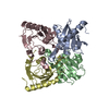
| |||||||||
|---|---|---|---|---|---|---|---|---|---|---|
| 1 |
| |||||||||
| Unit cell |
| |||||||||
| Components on special symmetry positions |
| |||||||||
| Details | CDD1 is a tetramer of identical subunits related by 222 symmetry. The asymmetric unit comprises 1 tetramer of chains A,B,C and D and includes four Zinc atoms. |
-Components
| #1: Protein | Mass: 15552.046 Da / Num. of mol.: 4 Source method: isolated from a genetically manipulated source Source: (gene. exp.) Gene: Cdd1p / Plasmid: pET28a / Production host:  #2: Chemical | ChemComp-ZN /   Mass: 65.409 Da / Num. of mol.: 4 / Source method: obtained synthetically / Formula: Zn Mass: 65.409 Da / Num. of mol.: 4 / Source method: obtained synthetically / Formula: Zn#3: Water | ChemComp-HOH / |  Mass: 18.015 Da / Num. of mol.: 318 / Source method: isolated from a natural source / Formula: H2O Mass: 18.015 Da / Num. of mol.: 318 / Source method: isolated from a natural source / Formula: H2O |
|---|
-Experimental details
-Experiment
| Experiment | Method: X-RAY DIFFRACTION / Number of used crystals: 1 |
|---|
- Sample preparation
| Crystal | Density Matthews: 2.13 Å3/Da / Density % sol: 42.22 % |
|---|---|
| Crystal grow | Temperature: 293 K / Method: vapor diffusion, hanging drop / pH: 5.5 Details: PEG 5000 MME, ammonium chloride, sodium succinate, DTT, sodium azide, pH 5.5, VAPOR DIFFUSION, HANGING DROP, temperature 293K |
-Data collection
| Diffraction |
| ||||||||||||
|---|---|---|---|---|---|---|---|---|---|---|---|---|---|
| Diffraction source | Source: SYNCHROTRON / Site: APS  / Beamline: 19-BM / Wavelength: 1.2574, 1.2831, 1.2828 / Beamline: 19-BM / Wavelength: 1.2574, 1.2831, 1.2828 | ||||||||||||
| Detector | Type: APS-1 / Detector: CCD / Date: Sep 17, 2001 / Details: Vertical Focusing Mirrors | ||||||||||||
| Radiation | Monochromator: Si-111 / Protocol: MAD / Monochromatic (M) / Laue (L): M / Scattering type: x-ray | ||||||||||||
| Radiation wavelength |
| ||||||||||||
| Reflection | Resolution: 2→29 Å / Num. all: 65372 / Num. obs: 63560 / % possible obs: 91.5 % / Observed criterion σ(F): 0 / Observed criterion σ(I): -3 / Redundancy: 4.5 % / Biso Wilson estimate: 29.7 Å2 / Rsym value: 0.064 / Net I/σ(I): 17.7 | ||||||||||||
| Reflection shell | Resolution: 2→2.07 Å / Redundancy: 4.5 % / Mean I/σ(I) obs: 2.1 / Num. unique all: 5597 / Rsym value: 0.313 / % possible all: 74.8 |
- Processing
| Software |
| ||||||||||||||||||||||||||||||||||||
|---|---|---|---|---|---|---|---|---|---|---|---|---|---|---|---|---|---|---|---|---|---|---|---|---|---|---|---|---|---|---|---|---|---|---|---|---|---|
| Refinement | Method to determine structure: MAD / Resolution: 2→29.05 Å / Rfactor Rfree error: 0.003 / Data cutoff high absF: 410906.6 / Data cutoff low absF: 0 / Isotropic thermal model: RESTRAINED / Cross valid method: THROUGHOUT / σ(F): 0 / Stereochemistry target values: Engh & Huber
| ||||||||||||||||||||||||||||||||||||
| Solvent computation | Solvent model: FLAT MODEL / Bsol: 41.9708 Å2 / ksol: 0.319008 e/Å3 | ||||||||||||||||||||||||||||||||||||
| Displacement parameters | Biso mean: 37.2 Å2
| ||||||||||||||||||||||||||||||||||||
| Refine analyze |
| ||||||||||||||||||||||||||||||||||||
| Refinement step | Cycle: LAST / Resolution: 2→29.05 Å
| ||||||||||||||||||||||||||||||||||||
| Refine LS restraints |
| ||||||||||||||||||||||||||||||||||||
| LS refinement shell | Resolution: 2→2.09 Å / Rfactor Rfree error: 0.012 / Total num. of bins used: 8
| ||||||||||||||||||||||||||||||||||||
| Xplor file |
|
