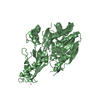
 Movie
Movie Controller


+ Open data

- Basic information
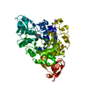
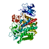
| Entry | Database: PDB / ID: 1af2 | |||||||||
|---|---|---|---|---|---|---|---|---|---|---|
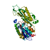
| Title | CRYSTAL STRUCTURE OF CYTIDINE DEAMINASE COMPLEXED WITH URIDINE | |||||||||
 Components Components | CYTIDINE DEAMINASE | |||||||||
 Keywords Keywords | HYDROLASE / DEAMINASE / PROTON TRANSFER / STRAIN / PRODUCT RELEASE | |||||||||
| Function / homology |  Function and homology information Function and homology informationpyrimidine nucleoside binding / deoxycytidine catabolic process / cytidine deaminase / cytidine deaminase activity / nucleobase-containing small molecule interconversion / protein homodimerization activity / zinc ion binding / identical protein binding / cytosol Similarity search - Function | |||||||||
| Biological species |  | |||||||||
| Method |  X-RAY DIFFRACTION / MOLECULAR REPLACEMENT / Resolution: 2.3 Å X-RAY DIFFRACTION / MOLECULAR REPLACEMENT / Resolution: 2.3 Å | |||||||||
 Authors Authors | Xiang, S. / Carter, C.W. | |||||||||
 Citation Citation | Journal: Biochemistry / Year: 1997 Title: The structure of the cytidine deaminase-product complex provides evidence for efficient proton transfer and ground-state destabilization. Authors: Xiang, S. / Short, S.A. / Wolfenden, R. / Carter Jr., C.W. | |||||||||
| History |
|
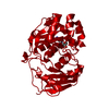
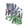
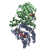
- Structure visualization
| Structure viewer | Molecule: MolmilJmol/JSmol |
|---|
- Downloads & links
-Download
| PDBx/mmCIF format | 1af2.cif.gz | 70.8 KB | Display | PDBx/mmCIF format |
|---|---|---|---|---|
| PDB format | pdb1af2.ent.gz | 51.7 KB | Display | PDB format |
| PDBx/mmJSON format | 1af2.json.gz | Tree view | PDBx/mmJSON format | |
| Others |  Other downloads Other downloads |
-Validation report
| Arichive directory | https://data.pdbj.org/pub/pdb/validation_reports/af/1af2ftp://data.pdbj.org/pub/pdb/validation_reports/af/1af2 | HTTPS FTP |
|---|
-Related structure data
| Related structure data |  1ctuS S: Starting model for refinement |
|---|---|
| Similar structure data |
-Links
 PDBj
PDBj

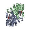
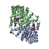
- Assembly
| Deposited unit | 
| ||||||||
|---|---|---|---|---|---|---|---|---|---|
| 1 | 
| ||||||||
| Unit cell |
|
-Components
| #1: Protein | Mass: 31569.785 Da / Num. of mol.: 1 Source method: isolated from a genetically manipulated source Source: (gene. exp.) |
|---|---|
| #2: Chemical | ChemComp-ZN /   Mass: 65.409 Da / Num. of mol.: 1 / Source method: obtained synthetically / Formula: Zn Mass: 65.409 Da / Num. of mol.: 1 / Source method: obtained synthetically / Formula: Zn |
| #3: Chemical | ChemComp-U5P /   Mass: 324.181 Da / Num. of mol.: 1 / Source method: obtained synthetically / Formula: C9H13N2O9P Mass: 324.181 Da / Num. of mol.: 1 / Source method: obtained synthetically / Formula: C9H13N2O9P |
| #4: Water | ChemComp-HOH /  Mass: 18.015 Da / Num. of mol.: 49 / Source method: isolated from a natural source / Formula: H2O Mass: 18.015 Da / Num. of mol.: 49 / Source method: isolated from a natural source / Formula: H2O |
| Has protein modification | N |
-Experimental details
-Experiment
| Experiment | Method: X-RAY DIFFRACTION / Number of used crystals: 1 |
|---|
- Sample preparation
| Crystal | Density Matthews: 5.187 Å3/Da / Density % sol: 76.289 % | |||||||||||||||||||||||||
|---|---|---|---|---|---|---|---|---|---|---|---|---|---|---|---|---|---|---|---|---|---|---|---|---|---|---|
| Crystal grow | pH: 6.2 / Details: pH 6.2 | |||||||||||||||||||||||||
| Crystal grow | *PLUS Temperature: 4 ℃ / Method: vapor diffusion, hanging drop | |||||||||||||||||||||||||
| Components of the solutions | *PLUS
|
-Data collection
| Diffraction | Mean temperature: 297 K |
|---|---|
| Diffraction source | Source: ROTATING ANODE / Type: RIGAKU RUH2R / Wavelength: 1.5418 |
| Detector | Type: RIGAKU RAXIS IIC / Detector: IMAGE PLATE / Date: Feb 1, 1993 / Details: MIRRORS |
| Radiation | Monochromator: NI FILTER / Monochromatic (M) / Laue (L): M / Scattering type: x-ray |
| Radiation wavelength | Wavelength: 1.5418 Å / Relative weight: 1 |
| Reflection | Resolution: 2.3→20 Å / Num. obs: 24137 / % possible obs: 80 % / Observed criterion σ(I): 1 / Redundancy: 2.7 % / Rmerge(I) obs: 0.075 / Rsym value: 0.08 / Net I/σ(I): 12 |
| Reflection shell | Resolution: 2.3→2.5 Å / Redundancy: 2.4 % / Rmerge(I) obs: 0.2 / Mean I/σ(I) obs: 2.5 / Rsym value: 0.22 / % possible all: 70 |
| Reflection | *PLUS Num. measured all: 65003 |
| Reflection shell | *PLUS % possible obs: 70 % |
- Processing
| Software |
| ||||||||||||||||||||
|---|---|---|---|---|---|---|---|---|---|---|---|---|---|---|---|---|---|---|---|---|---|
| Refinement | Method to determine structure: MOLECULAR REPLACEMENT Starting model: PDB ENTRY 1CTU Resolution: 2.3→7 Å / Data cutoff high absF: 10000000 / Data cutoff low absF: 0.001 / σ(F): 2 /
| ||||||||||||||||||||
| Refinement step | Cycle: LAST / Resolution: 2.3→7 Å
| ||||||||||||||||||||
| Xplor file |
| ||||||||||||||||||||
| Software | *PLUS Name: X-PLOR / Version: 3 / Classification: refinement | ||||||||||||||||||||
| Refinement | *PLUS Rfactor obs: 0.19 | ||||||||||||||||||||
| Solvent computation | *PLUS | ||||||||||||||||||||
| Displacement parameters | *PLUS | ||||||||||||||||||||
| Refine LS restraints | *PLUS
|
