 Movie
Movie Controller

[English] 日本語
 Yorodumi
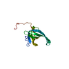
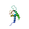
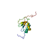
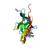
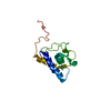
- PDB-1r57: NMR Solution Structure of a GCN5-like putative N-acetyltransferas... -
+ Open data

- Basic information
| Entry | Database: PDB / ID: 1r57 | |||||||||
|---|---|---|---|---|---|---|---|---|---|---|
| Title | NMR Solution Structure of a GCN5-like putative N-acetyltransferase from Staphylococcus aureus. Northeast Structural Genomics Consortium Target ZR31 | |||||||||
 Components Components | conserved hypothetical protein | |||||||||
 Keywords Keywords | TRANSFERASE / GCN5 / N-acetyltransferase / Structural genomics / PSI / Protein Structure Initiative / Northeast Structural Genomics Consortium / NESG | |||||||||
| Function / homology |  Function and homology information Function and homology informationacyltransferase activity, transferring groups other than amino-acyl groups Similarity search - Function | |||||||||
| Biological species |   Staphylococcus aureus (bacteria) Staphylococcus aureus (bacteria) | |||||||||
| Method | SOLUTION NMR / automated generation of initial distance restraint set distance geometry simulated annealing final refinement in explicit solvent | |||||||||
 Authors Authors | Cort, J.R. / Acton, T.B. / Ma, L. / Xiao, R.B. / Montelione, G.T. / Kennedy, M.A. / Northeast Structural Genomics Consortium (NESG) | |||||||||
 Citation Citation | Journal: J.STRUCT.FUNCT.GENOM. / Year: 2008 Title: Structure of an acetyl-CoA binding protein from Staphylococcus aureus representing a novel subfamily of GCN5-related N-acetyltransferase-like proteins. Authors: Cort, J.R. / Ramelot, T.A. / Murray, D. / Acton, T.B. / Ma, L.C. / Xiao, R. / Montelione, G.T. / Kennedy, M.A. | |||||||||
| History |
| |||||||||
| Remark 999 | sequence The protein that is the subject of this structure determination was cloned from ...sequence The protein that is the subject of this structure determination was cloned from Staphylococcus aureus subsp. Rosenbach. This strain has not been the subject of genome sequencing, however three other strains have been sequenced: MW2, Mu50,and N315. The amino acid residues at three positions in the protein structure described here differ from those at some or all of the same positions in the sequences from the three sequenced strains. These mutations, indicated by the notation XnY where the X is the residue in the sequenced strain, n is the position number, and Y is the residue in the structure described here, are as follows: Strain MW2: V57L and H68N Strains Mu50 and N315: H21N, V57L, and H68N |
- Structure visualization
| Structure viewer | Molecule: MolmilJmol/JSmol |
|---|
- Downloads & links
-Download
| PDBx/mmCIF format | 1r57.cif.gz | 622.9 KB | Display | PDBx/mmCIF format |
|---|---|---|---|---|
| PDB format | pdb1r57.ent.gz | 521.7 KB | Display | PDB format |
| PDBx/mmJSON format | 1r57.json.gz | Tree view | PDBx/mmJSON format | |
| Others |  Other downloads Other downloads |
-Validation report
| Arichive directory | https://data.pdbj.org/pub/pdb/validation_reports/r5/1r57ftp://data.pdbj.org/pub/pdb/validation_reports/r5/1r57 | HTTPS FTP |
|---|
-Related structure data
| Related structure data |  2h5mC C: citing same article ( |
|---|---|
| Similar structure data | |
| Other databases |
|


-Links
 PDBj
PDBj

- Assembly
| Deposited unit | 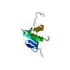
| |||||||||
|---|---|---|---|---|---|---|---|---|---|---|
| 1 |
| |||||||||
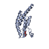
| NMR ensembles |
|
-Components
| #1: Protein | Mass: 11631.878 Da / Num. of mol.: 1 Source method: isolated from a genetically manipulated source Source: (gene. exp.) Staphylococcus aureus (bacteria) / Gene: SA2309 or MW2441 or SAV2521 / Plasmid: pET21 / Species (production host): Escherichia coli / Production host: References: UniProt: Q99RB4, UniProt: A0A0H3JTC0*PLUS, Transferases; Acyltransferases; Transferring groups other than aminoacyl groups |
|---|
-Experimental details
-Experiment
| Experiment | Method: SOLUTION NMR | ||||||||||||||||||||||||
|---|---|---|---|---|---|---|---|---|---|---|---|---|---|---|---|---|---|---|---|---|---|---|---|---|---|
| NMR experiment |
| ||||||||||||||||||||||||
| NMR details | Text: amide proton exchange was measured by dissolving a lyophilized protonated sample in D2O |
- Sample preparation
| Details |
| ||||||||||||
|---|---|---|---|---|---|---|---|---|---|---|---|---|---|
| Sample conditions | Ionic strength: 0.115 / pH: 6.5 / Pressure: ambient / Temperature: 293 K | ||||||||||||
| Crystal grow | *PLUS Method: other / Details: NMR |
-NMR measurement
| NMR spectrometer |
|
|---|
- Processing
| NMR software |
| ||||||||||||||||||||||||||||
|---|---|---|---|---|---|---|---|---|---|---|---|---|---|---|---|---|---|---|---|---|---|---|---|---|---|---|---|---|---|
| Refinement | Method: automated generation of initial distance restraint set distance geometry simulated annealing final refinement in explicit solvent Software ordinal: 1 Details: Backbone and sidechain assignments were determined manually from triple-resonance NMR data. NOE distance restraints were derived automatically from peak-picked data with AutoStructure, then ...Details: Backbone and sidechain assignments were determined manually from triple-resonance NMR data. NOE distance restraints were derived automatically from peak-picked data with AutoStructure, then error-checked and corrected manually. The structure is based on 848 restraints: 710 meaningful distance restraints, 58 hydrogen bond restraints, and 80 dihedral angle restraints. There are 9.5 restraints per restrained residue. Phi dihedral restraints were derived from the HNHA experiment and TALOS. Psi dihedral restraints were derived from NOE ratios, secondary structure propensities evident in preliminary structures, alpha carbon chemical shifts, and TALOS. Residues 44-51 comprise a poorly-defined loop in this ensemble of structures. Residues 1-3 and 94-102 are unstructured termini. | ||||||||||||||||||||||||||||
| NMR representative | Selection criteria: similarity to average, few violations, and low energy | ||||||||||||||||||||||||||||
| NMR ensemble | Conformer selection criteria: structures with fewest restraint violations and lowest energy Conformers calculated total number: 30 / Conformers submitted total number: 20 |
