 Movie
Movie Controller

[English] 日本語
 Yorodumi
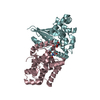
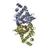
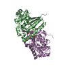
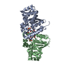
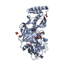
- PDB-1r4w: Crystal structure of Mitochondrial class kappa glutathione transferase -
+ Open data

- Basic information
| Entry | Database: PDB / ID: 1r4w | ||||||
|---|---|---|---|---|---|---|---|
| Title | Crystal structure of Mitochondrial class kappa glutathione transferase | ||||||
 Components Components | Glutathione S-transferase, mitochondrial | ||||||
 Keywords Keywords | TRANSFERASE / glutathione-S-transferase / glutathione transferase / kappa GST / rGSTK1-1 | ||||||
| Function / homology |  Function and homology information Function and homology informationPeroxisomal protein import / Glutathione conjugation / glutathione peroxidase activity / glutathione transferase / glutathione transferase activity / epithelial cell differentiation / glutathione metabolic process / peroxisome / mitochondrial matrix / mitochondrion Similarity search - Function | ||||||
| Biological species |  | ||||||
| Method |  X-RAY DIFFRACTION / SYNCHROTRON / SIR with anomalous Hg / Resolution: 2.5 Å X-RAY DIFFRACTION / SYNCHROTRON / SIR with anomalous Hg / Resolution: 2.5 Å | ||||||
 Authors Authors | Ladner, J.E. / Parsons, J.F. / Rife, C.L. / Gilliland, G.L. / Armstrong, R.N. | ||||||
 Citation Citation | Journal: Biochemistry / Year: 2004 Title: Parallel Evolutionary Pathways for Glutathione Transferases: Structure and Mechanism of the Mitochondrial Class Kappa Enzyme rGSTK1-1 Authors: Ladner, J.E. / Parsons, J.F. / Rife, C.L. / Gilliland, G.L. / Armstrong, R.N. | ||||||
| History |
|
- Structure visualization
| Structure viewer | Molecule: MolmilJmol/JSmol |
|---|
- Downloads & links
-Download
| PDBx/mmCIF format | 1r4w.cif.gz | 185.7 KB | Display | PDBx/mmCIF format |
|---|---|---|---|---|
| PDB format | pdb1r4w.ent.gz | 150.1 KB | Display | PDB format |
| PDBx/mmJSON format | 1r4w.json.gz | Tree view | PDBx/mmJSON format | |
| Others |  Other downloads Other downloads |
-Validation report
| Arichive directory | https://data.pdbj.org/pub/pdb/validation_reports/r4/1r4wftp://data.pdbj.org/pub/pdb/validation_reports/r4/1r4w | HTTPS FTP |
|---|
-Related structure data
| Similar structure data |
|---|


-Links
 PDBj
PDBj

- Assembly
| Deposited unit | 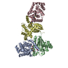
| ||||||||
|---|---|---|---|---|---|---|---|---|---|
| 1 | 
| ||||||||
| 2 | 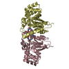
| ||||||||
| Unit cell |
|
-Components
| #1: Protein | Mass: 25524.969 Da / Num. of mol.: 4 Source method: isolated from a genetically manipulated source Source: (gene. exp.)  #2: Chemical | ChemComp-GSH /   Mass: 307.323 Da / Num. of mol.: 4 / Source method: obtained synthetically / Formula: C10H17N3O6S Mass: 307.323 Da / Num. of mol.: 4 / Source method: obtained synthetically / Formula: C10H17N3O6S#3: Water | ChemComp-HOH / |  Mass: 18.015 Da / Num. of mol.: 266 / Source method: isolated from a natural source / Formula: H2O Mass: 18.015 Da / Num. of mol.: 266 / Source method: isolated from a natural source / Formula: H2ONonpolymer details | HOH 723 MAY BE A PARTIALLY OCCUPIED SULFATE ION. | |
|---|
-Experimental details
-Experiment
| Experiment | Method: X-RAY DIFFRACTION / Number of used crystals: 1 |
|---|
- Sample preparation
| Crystal | Density Matthews: 2.61 Å3/Da / Density % sol: 52.92 % | |||||||||||||||||||||||||||||||||||||||||||||||||
|---|---|---|---|---|---|---|---|---|---|---|---|---|---|---|---|---|---|---|---|---|---|---|---|---|---|---|---|---|---|---|---|---|---|---|---|---|---|---|---|---|---|---|---|---|---|---|---|---|---|---|
| Crystal grow | Temperature: 277 K / Method: vapor diffusion, hanging drop / pH: 4 Details: well: 13-17% (w/v) PEG 2K, 40-80 mM lithium sulfate, 100 mM sodium citrate; drop: 1:1 protein:well, 0.05% beta-octyl-glucopyranoside, 1.5 mM glutathione, pH 4.0, VAPOR DIFFUSION, HANGING DROP, temperature 277K | |||||||||||||||||||||||||||||||||||||||||||||||||
| Crystal grow | *PLUS Temperature: 4 ℃ / Method: vapor diffusion, hanging drop / PH range low: 4.4 / PH range high: 4 | |||||||||||||||||||||||||||||||||||||||||||||||||
| Components of the solutions | *PLUS
|
-Data collection
| Diffraction | Mean temperature: 100 K |
|---|---|
| Diffraction source | Source: SYNCHROTRON / Site: APS  / Beamline: 14-BM-D / Wavelength: 1 Å / Beamline: 14-BM-D / Wavelength: 1 Å |
| Radiation | Protocol: SINGLE WAVELENGTH / Monochromatic (M) / Laue (L): M / Scattering type: x-ray |
| Radiation wavelength | Wavelength: 1 Å / Relative weight: 1 |
| Reflection | Resolution: 2.5→30 Å / Num. all: 35795 / Num. obs: 35795 / % possible obs: 98.4 % / Redundancy: 4 % / Biso Wilson estimate: 33.9 Å2 / Rsym value: 0.072 / Net I/σ(I): 11 |
| Reflection shell | Resolution: 2.5→2.59 Å / Redundancy: 3 % / Rmerge(I) obs: 0.166 / Mean I/σ(I) obs: 5 / % possible all: 88.7 |
| Reflection | *PLUS Highest resolution: 2.5 Å / Num. measured all: 115187 / Rmerge(I) obs: 0.072 |
| Reflection shell | *PLUS % possible obs: 88.7 % / Mean I/σ(I) obs: 5 |
- Processing
| Software |
| |||||||||||||||||||||||||
|---|---|---|---|---|---|---|---|---|---|---|---|---|---|---|---|---|---|---|---|---|---|---|---|---|---|---|
| Refinement | Method to determine structure: SIR with anomalous Hg / Resolution: 2.5→30 Å / Cross valid method: THROUGHOUT / σ(F): 0 / Stereochemistry target values: Engh & Huber / Details: Freidel pairs were used in the refinement.
| |||||||||||||||||||||||||
| Refine analyze |
| |||||||||||||||||||||||||
| Refinement step | Cycle: LAST / Resolution: 2.5→30 Å
| |||||||||||||||||||||||||
| Refine LS restraints |
| |||||||||||||||||||||||||
| Refinement | *PLUS Highest resolution: 2.5 Å / Lowest resolution: 30 Å | |||||||||||||||||||||||||
| Solvent computation | *PLUS | |||||||||||||||||||||||||
| Displacement parameters | *PLUS |
