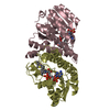
 Movie
Movie Controller


+ Open data

- Basic information
| Entry | Database: PDB / ID: 2z1k | |||||||||
|---|---|---|---|---|---|---|---|---|---|---|
| Title | Crystal Structure of Ttha1563 from Thermus thermophilus HB8 | |||||||||
 Components Components | (Neo)pullulanase | |||||||||
 Keywords Keywords | HYDROLASE / Structural Genomics / NPPSFA / National Project on Protein Structural and Functional Analyses / RIKEN Structural Genomics/Proteomics Initiative / RSGI | |||||||||
| Function / homology |  Function and homology information Function and homology informationhydrolase activity, acting on glycosyl bonds / carbohydrate metabolic process Similarity search - Function | |||||||||
| Biological species |   Thermus thermophilus (bacteria) Thermus thermophilus (bacteria) | |||||||||
| Method |  X-RAY DIFFRACTION / SYNCHROTRON / MOLECULAR REPLACEMENT / Resolution: 2.3 Å X-RAY DIFFRACTION / SYNCHROTRON / MOLECULAR REPLACEMENT / Resolution: 2.3 Å | |||||||||
 Authors Authors | Niwa, H. / Shimada, A. / Matsunaga, E. / Kuramitsu, S. / Yokoyama, S. / RIKEN Structural Genomics/Proteomics Initiative (RSGI) | |||||||||
 Citation Citation | Journal: To be Published Title: Crystal Structure of Ttha1563 from Thermus thermophilus HB8 Authors: Niwa, H. / Shimada, A. / Matsunaga, E. / Kuramitsu, S. / Yokoyama, S. | |||||||||
| History |
|
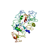
- Structure visualization
| Structure viewer | Molecule: MolmilJmol/JSmol |
|---|
- Downloads & links
-Download
| PDBx/mmCIF format | 2z1k.cif.gz | 402.7 KB | Display | PDBx/mmCIF format |
|---|---|---|---|---|
| PDB format | pdb2z1k.ent.gz | 332.7 KB | Display | PDB format |
| PDBx/mmJSON format | 2z1k.json.gz | Tree view | PDBx/mmJSON format | |
| Others |  Other downloads Other downloads |
-Validation report
| Arichive directory | https://data.pdbj.org/pub/pdb/validation_reports/z1/2z1kftp://data.pdbj.org/pub/pdb/validation_reports/z1/2z1k | HTTPS FTP |
|---|
-Related structure data
| Related structure data |  1j0hS S: Starting model for refinement |
|---|---|
| Similar structure data | |
| Other databases |

-Links
 PDBj
PDBj


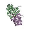
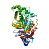
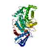
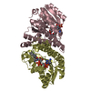
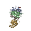
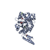
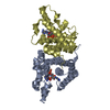
- Assembly
| Deposited unit | 
| ||||||||
|---|---|---|---|---|---|---|---|---|---|
| 1 | 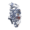
| ||||||||
| 2 | 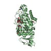
| ||||||||
| 3 | 
| ||||||||
| 4 | 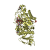
| ||||||||
| 5 | 
| ||||||||
| 6 | 
| ||||||||
| Unit cell |
|
-Components
| #1: Protein | ( Mass: 54200.363 Da / Num. of mol.: 4 Source method: isolated from a genetically manipulated source Source: (gene. exp.) Thermus thermophilus (bacteria) / Strain: HB8 / Plasmid: plasmid / Production host: #2: Polysaccharide | Cycloheptakis-(1-4)-(alpha-D-glucopyranose) / beta-cyclodextrin 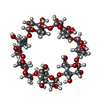  Source method: isolated from a genetically manipulated source Details: cyclic oligosaccharide / References: beta-cyclodextrin #3: Polysaccharide | alpha-D-glucopyranose-(1-4)-alpha-D-glucopyranose-(1-4)-alpha-D-glucopyranose / alpha-maltotriose   Source method: isolated from a genetically manipulated source Details: oligosaccharide / References: alpha-maltotriose #4: Chemical | ChemComp-PO4 / |   Mass: 94.971 Da / Num. of mol.: 1 / Source method: obtained synthetically / Formula: PO4 Mass: 94.971 Da / Num. of mol.: 1 / Source method: obtained synthetically / Formula: PO4#5: Water | ChemComp-HOH / |  Mass: 18.015 Da / Num. of mol.: 781 / Source method: isolated from a natural source / Formula: H2O Mass: 18.015 Da / Num. of mol.: 781 / Source method: isolated from a natural source / Formula: H2OHas protein modification | Y | |
|---|
-Experimental details
-Experiment
| Experiment | Method: X-RAY DIFFRACTION / Number of used crystals: 1 |
|---|
- Sample preparation
| Crystal | Density Matthews: 2.08 Å3/Da / Density % sol: 40.91 % |
|---|---|
| Crystal grow | Temperature: 293 K / Method: vapor diffusion, hanging drop / pH: 4.6 Details: 0.3M NH4H2PO4, 20% PEG 3350, 40% ETHANOL, pH 4.60, VAPOR DIFFUSION, HANGING DROP, temperature 293K |
-Data collection
| Diffraction | Mean temperature: 100 K |
|---|---|
| Diffraction source | Source: SYNCHROTRON / Site: SPring-8  / Beamline: BL44B2 / Wavelength: 1 Å / Beamline: BL44B2 / Wavelength: 1 Å |
| Detector | Type: ADSC QUANTUM 210 / Detector: CCD / Date: Nov 19, 2006 |
| Radiation | Protocol: SINGLE WAVELENGTH / Monochromatic (M) / Laue (L): M / Scattering type: x-ray |
| Radiation wavelength | Wavelength: 1 Å / Relative weight: 1 |
| Reflection | Resolution: 2.3→50 Å / Num. obs: 79591 / % possible obs: 100 % / Observed criterion σ(I): 0 / Redundancy: 7.6 % / Biso Wilson estimate: 31.4 Å2 / Rmerge(I) obs: 0.076 / Net I/σ(I): 28.1 |
| Reflection shell | Resolution: 2.3→2.38 Å / Redundancy: 7.6 % / Rmerge(I) obs: 0.38 / Mean I/σ(I) obs: 5.6 / % possible all: 100 |
- Processing
| Software |
| ||||||||||||||||||||||||||||||||||||||||||||||||||||||||||||||||||||||||||||||||
|---|---|---|---|---|---|---|---|---|---|---|---|---|---|---|---|---|---|---|---|---|---|---|---|---|---|---|---|---|---|---|---|---|---|---|---|---|---|---|---|---|---|---|---|---|---|---|---|---|---|---|---|---|---|---|---|---|---|---|---|---|---|---|---|---|---|---|---|---|---|---|---|---|---|---|---|---|---|---|---|---|---|
| Refinement | Method to determine structure: MOLECULAR REPLACEMENT Starting model: PDB ENTRY 1J0H Resolution: 2.3→46.66 Å / Rfactor Rfree error: 0.004 / Data cutoff high absF: 2709718.86 / Data cutoff low absF: 0 / Isotropic thermal model: RESTRAINED / Cross valid method: THROUGHOUT / σ(F): 0 / Stereochemistry target values: ENGH & HUBER
| ||||||||||||||||||||||||||||||||||||||||||||||||||||||||||||||||||||||||||||||||
| Solvent computation | Solvent model: FLAT MODEL / Bsol: 36.96 Å2 / ksol: 0.36 e/Å3 | ||||||||||||||||||||||||||||||||||||||||||||||||||||||||||||||||||||||||||||||||
| Displacement parameters | Biso mean: 30.9 Å2
| ||||||||||||||||||||||||||||||||||||||||||||||||||||||||||||||||||||||||||||||||
| Refine analyze |
| ||||||||||||||||||||||||||||||||||||||||||||||||||||||||||||||||||||||||||||||||
| Refinement step | Cycle: LAST / Resolution: 2.3→46.66 Å
| ||||||||||||||||||||||||||||||||||||||||||||||||||||||||||||||||||||||||||||||||
| Refine LS restraints |
| ||||||||||||||||||||||||||||||||||||||||||||||||||||||||||||||||||||||||||||||||
| LS refinement shell | Resolution: 2.3→2.44 Å / Rfactor Rfree error: 0.011 / Total num. of bins used: 6
| ||||||||||||||||||||||||||||||||||||||||||||||||||||||||||||||||||||||||||||||||
| Xplor file |
|
