 Movie
Movie Controller

[English] 日本語
 Yorodumi
- PDB-1r3w: Uroporphyrinogen Decarboxylase Y164F mutant in complex with copro... -
+ Open data

- Basic information
| Entry | Database: PDB / ID: 1r3w | ||||||
|---|---|---|---|---|---|---|---|
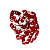
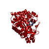
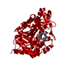
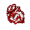
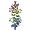
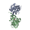
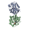
| Title | Uroporphyrinogen Decarboxylase Y164F mutant in complex with coproporphyrinogen-III | ||||||
 Components Components | Uroporphyrinogen Decarboxylase | ||||||
 Keywords Keywords | LYASE / uroporphyrinogen decarboxylase coproporphyrinogen / X-ray crystallography | ||||||
| Function / homology |  Function and homology information Function and homology informationporphyrin-containing compound catabolic process / uroporphyrinogen decarboxylase / uroporphyrinogen decarboxylase activity / porphyrin-containing compound metabolic process / heme A biosynthetic process / heme B biosynthetic process / : / Heme biosynthesis / heme biosynthetic process / nucleoplasm / cytosol Similarity search - Function | ||||||
| Biological species |  Homo sapiens (human) Homo sapiens (human) | ||||||
| Method |  X-RAY DIFFRACTION / FOURIER SYNTHESIS / Resolution: 1.7 Å X-RAY DIFFRACTION / FOURIER SYNTHESIS / Resolution: 1.7 Å | ||||||
 Authors Authors | Phillips, J.D. / Whitby, F.G. / Kushner, J.P. / Hill, C.P. | ||||||
 Citation Citation | Journal: Embo J. / Year: 2003 Title: Structural basis for tetrapyrrole coordination by uroporphyrinogen decarboxylase Authors: Phillips, J.D. / Whitby, F.G. / Kushner, J.P. / Hill, C.P. | ||||||
| History |
|
- Structure visualization
| Structure viewer | Molecule: MolmilJmol/JSmol |
|---|
- Downloads & links
-Download
| PDBx/mmCIF format | 1r3w.cif.gz | 95.4 KB | Display | PDBx/mmCIF format |
|---|---|---|---|---|
| PDB format | pdb1r3w.ent.gz | 72.1 KB | Display | PDB format |
| PDBx/mmJSON format | 1r3w.json.gz | Tree view | PDBx/mmJSON format | |
| Others |  Other downloads Other downloads |
-Validation report
| Arichive directory | https://data.pdbj.org/pub/pdb/validation_reports/r3/1r3wftp://data.pdbj.org/pub/pdb/validation_reports/r3/1r3w | HTTPS FTP |
|---|
-Related structure data
| Related structure data |  1r3qC  1r3rC  1r3sC  1r3tC  1r3vC  1r3yC  1uroS S: Starting model for refinement C: citing same article ( |
|---|---|
| Similar structure data |


-Links
 PDBj
PDBj

- Assembly
| Deposited unit | 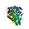
| ||||||||||
|---|---|---|---|---|---|---|---|---|---|---|---|
| 1 | 
| ||||||||||
| Unit cell |
| ||||||||||
| Details | URO-D is a dimer in solution and in the crystal. The dimer is formed by the 2-fold crystallographic rotation |
-Components
| #1: Protein | Mass: 40815.766 Da / Num. of mol.: 1 / Mutation: Y164F Source method: isolated from a genetically manipulated source Source: (gene. exp.) Homo sapiens (human) / Production host:  |
|---|---|
| #2: Chemical | ChemComp-CP3 /   Mass: 660.757 Da / Num. of mol.: 1 / Source method: obtained synthetically / Formula: C36H44N4O8 Mass: 660.757 Da / Num. of mol.: 1 / Source method: obtained synthetically / Formula: C36H44N4O8 |
| #3: Water | ChemComp-HOH /  Mass: 18.015 Da / Num. of mol.: 354 / Source method: isolated from a natural source / Formula: H2O Mass: 18.015 Da / Num. of mol.: 354 / Source method: isolated from a natural source / Formula: H2O |
-Experimental details
-Experiment
| Experiment | Method: X-RAY DIFFRACTION / Number of used crystals: 1 |
|---|
- Sample preparation
| Crystal | Density Matthews: 2.4 Å3/Da / Density % sol: 48.4 % |
|---|---|
| Crystal grow | Temperature: 294 K / Method: liquid diffusion / pH: 6.5 Details: PBG, PBG-D and U3S were added to the protein solution in an anaerobic chamber. URO-D was active under these conditions, converting uro'gen-III to cop'gen-III. 1.5 M citrate, pH 6.5, LIQUID ...Details: PBG, PBG-D and U3S were added to the protein solution in an anaerobic chamber. URO-D was active under these conditions, converting uro'gen-III to cop'gen-III. 1.5 M citrate, pH 6.5, LIQUID DIFFUSION, temperature 294K |
-Data collection
| Diffraction | Mean temperature: 100 K |
|---|---|
| Diffraction source | Source: ROTATING ANODE / Type: RIGAKU RU200 / Wavelength: 1.5418 Å |
| Detector | Type: RIGAKU RAXIS IV / Detector: IMAGE PLATE / Date: Jun 15, 2003 |
| Radiation | Protocol: SINGLE WAVELENGTH / Monochromatic (M) / Laue (L): M / Scattering type: x-ray |
| Radiation wavelength | Wavelength: 1.5418 Å / Relative weight: 1 |
| Reflection | Resolution: 1.7→87.71 Å / Num. all: 49087 / Num. obs: 49087 / % possible obs: 99 % / Observed criterion σ(F): 0 / Observed criterion σ(I): 0 / Redundancy: 5.9 % / Rmerge(I) obs: 0.071 / Rsym value: 0.071 / Net I/σ(I): 12 |
| Reflection shell | Resolution: 1.7→1.76 Å / Redundancy: 5.9 % / Rmerge(I) obs: 0.39 / Mean I/σ(I) obs: 1.8 / Rsym value: 0.39 / % possible all: 99 |
- Processing
| Software |
| ||||||||||||||||||||||||||||||||||||||||||||||||||||||||||||||||||||||||||||||||||||||||||||||||||||||||||||||||||||||||||||||||||
|---|---|---|---|---|---|---|---|---|---|---|---|---|---|---|---|---|---|---|---|---|---|---|---|---|---|---|---|---|---|---|---|---|---|---|---|---|---|---|---|---|---|---|---|---|---|---|---|---|---|---|---|---|---|---|---|---|---|---|---|---|---|---|---|---|---|---|---|---|---|---|---|---|---|---|---|---|---|---|---|---|---|---|---|---|---|---|---|---|---|---|---|---|---|---|---|---|---|---|---|---|---|---|---|---|---|---|---|---|---|---|---|---|---|---|---|---|---|---|---|---|---|---|---|---|---|---|---|---|---|---|---|
| Refinement | Method to determine structure: FOURIER SYNTHESIS Starting model: 1URO Resolution: 1.7→87.71 Å / Cor.coef. Fo:Fc: 0.969 / Cor.coef. Fo:Fc free: 0.958 / SU B: 1.78 / SU ML: 0.058 / Cross valid method: THROUGHOUT / σ(F): 0 / ESU R: 0.085 / ESU R Free: 0.086 / Stereochemistry target values: MAXIMUM LIKELIHOOD / Details: HYDROGENS HAVE BEEN ADDED IN THE RIDING POSITIONS
| ||||||||||||||||||||||||||||||||||||||||||||||||||||||||||||||||||||||||||||||||||||||||||||||||||||||||||||||||||||||||||||||||||
| Solvent computation | Ion probe radii: 0.8 Å / Shrinkage radii: 0.8 Å / VDW probe radii: 1.4 Å / Solvent model: BABINET MODEL WITH MASK | ||||||||||||||||||||||||||||||||||||||||||||||||||||||||||||||||||||||||||||||||||||||||||||||||||||||||||||||||||||||||||||||||||
| Displacement parameters | Biso mean: 23.551 Å2
| ||||||||||||||||||||||||||||||||||||||||||||||||||||||||||||||||||||||||||||||||||||||||||||||||||||||||||||||||||||||||||||||||||
| Refinement step | Cycle: LAST / Resolution: 1.7→87.71 Å
| ||||||||||||||||||||||||||||||||||||||||||||||||||||||||||||||||||||||||||||||||||||||||||||||||||||||||||||||||||||||||||||||||||
| Refine LS restraints |
| ||||||||||||||||||||||||||||||||||||||||||||||||||||||||||||||||||||||||||||||||||||||||||||||||||||||||||||||||||||||||||||||||||
| LS refinement shell | Resolution: 1.7→1.794 Å / Total num. of bins used: 10 /
|
