 Movie
Movie Controller


+ Open data

- Basic information
| Entry | Database: PDB / ID: 1r3r | ||||||
|---|---|---|---|---|---|---|---|
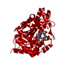
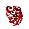
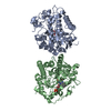
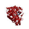
| Title | Uroporphyrinogen Decarboxylase with mutation D86N | ||||||
 Components Components | Uroporphyrinogen Decarboxylase | ||||||
 Keywords Keywords | LYASE / uroporphyrinogen decarboxylase coproporphyrinogen / X-ray crystallography | ||||||
| Function / homology |  Function and homology information Function and homology informationporphyrin-containing compound catabolic process / uroporphyrinogen decarboxylase / uroporphyrinogen decarboxylase activity / porphyrin-containing compound metabolic process / heme A biosynthetic process / heme B biosynthetic process / : / Heme biosynthesis / heme biosynthetic process / nucleoplasm / cytosol Similarity search - Function | ||||||
| Biological species |  Homo sapiens (human) Homo sapiens (human) | ||||||
| Method |  X-RAY DIFFRACTION / FOURIER SYNTHESIS / Resolution: 1.85 Å X-RAY DIFFRACTION / FOURIER SYNTHESIS / Resolution: 1.85 Å | ||||||
 Authors Authors | Phillips, J.D. / Whitby, F.G. / Kushner, J.P. / Hill, C.P. | ||||||
 Citation Citation | Journal: Embo J. / Year: 2003 Title: Structural basis for tetrapyrrole coordination by uroporphyrinogen decarboxylase Authors: Phillips, J.D. / Whitby, F.G. / Kushner, J.P. / Hill, C.P. | ||||||
| History |
|
- Structure visualization
| Structure viewer | Molecule: MolmilJmol/JSmol |
|---|
- Downloads & links
-Download
| PDBx/mmCIF format | 1r3r.cif.gz | 95 KB | Display | PDBx/mmCIF format |
|---|---|---|---|---|
| PDB format | pdb1r3r.ent.gz | 72.4 KB | Display | PDB format |
| PDBx/mmJSON format | 1r3r.json.gz | Tree view | PDBx/mmJSON format | |
| Others |  Other downloads Other downloads |
-Validation report
| Arichive directory | https://data.pdbj.org/pub/pdb/validation_reports/r3/1r3rftp://data.pdbj.org/pub/pdb/validation_reports/r3/1r3r | HTTPS FTP |
|---|
-Related structure data
| Related structure data |  1r3qC  1r3sC  1r3tC  1r3vC  1r3wC  1r3yC  1uroS S: Starting model for refinement C: citing same article ( |
|---|---|
| Similar structure data |


-Links
 PDBj
PDBj

- Assembly
| Deposited unit | 
| ||||||||||
|---|---|---|---|---|---|---|---|---|---|---|---|
| 1 | 
| ||||||||||
| Unit cell |
| ||||||||||
| Details | One URO-D monomer forms the asymmetric unit. URO-D is a dimer in solution and in the crystal. The dimer is formed by the 2-fold crystallographic rotation. |
-Components
| #1: Protein | Mass: 40830.781 Da / Num. of mol.: 1 / Mutation: D86N Source method: isolated from a genetically manipulated source Source: (gene. exp.) Homo sapiens (human) / Production host:  |
|---|---|
| #2: Water | ChemComp-HOH /  Mass: 18.015 Da / Num. of mol.: 381 / Source method: isolated from a natural source / Formula: H2O Mass: 18.015 Da / Num. of mol.: 381 / Source method: isolated from a natural source / Formula: H2O |
-Experimental details
-Experiment
| Experiment | Method: X-RAY DIFFRACTION / Number of used crystals: 1 |
|---|
- Sample preparation
| Crystal | Density Matthews: 2.37 Å3/Da / Density % sol: 47.7 % | ||||||||||||||||||||||||||||||||||||||||||
|---|---|---|---|---|---|---|---|---|---|---|---|---|---|---|---|---|---|---|---|---|---|---|---|---|---|---|---|---|---|---|---|---|---|---|---|---|---|---|---|---|---|---|---|
| Crystal grow | Temperature: 294 K / Method: liquid diffusion / pH: 6.5 Details: The crystal trial was conducted in an anaerobic chamber, with PBG, PBG-D added to the solution. These conditions are conducive to formation of an enzyme product complex, as the enzyme is ...Details: The crystal trial was conducted in an anaerobic chamber, with PBG, PBG-D added to the solution. These conditions are conducive to formation of an enzyme product complex, as the enzyme is typically active under these conditions (see related strcuture 1R3Q). There is not a ligand bound in this structure. 1.5 M citrate, pH 6.5, LIQUID DIFFUSION, temperature 294K | ||||||||||||||||||||||||||||||||||||||||||
| Crystal grow | *PLUS Temperature: 21 ℃ / pH: 7.5 / Method: vapor diffusion, sitting drop / Details: Phillips, J.D., (1997) Protein Sci., 6, 1343. | ||||||||||||||||||||||||||||||||||||||||||
| Components of the solutions | *PLUS
|
-Data collection
| Diffraction | Mean temperature: 100 K |
|---|---|
| Diffraction source | Source: ROTATING ANODE / Type: RIGAKU RU200 / Wavelength: 1.5418 Å |
| Detector | Type: RIGAKU RAXIS IIC / Detector: IMAGE PLATE / Date: Jun 15, 2003 |
| Radiation | Protocol: SINGLE WAVELENGTH / Monochromatic (M) / Laue (L): M / Scattering type: x-ray |
| Radiation wavelength | Wavelength: 1.5418 Å / Relative weight: 1 |
| Reflection | Resolution: 1.85→87.71 Å / Num. all: 36928 / Num. obs: 36928 / % possible obs: 96 % / Observed criterion σ(F): 0 / Observed criterion σ(I): 0 / Redundancy: 5 % / Rmerge(I) obs: 0.083 / Rsym value: 0.083 / Net I/σ(I): 9 |
| Reflection shell | Resolution: 1.85→1.92 Å / Redundancy: 5 % / Rmerge(I) obs: 0.437 / Mean I/σ(I) obs: 2.8 / Rsym value: 0.437 / % possible all: 80 |
| Reflection | *PLUS Lowest resolution: 20 Å / % possible obs: 96 % / Num. measured all: 175858 |
| Reflection shell | *PLUS % possible obs: 79.8 % / Mean I/σ(I) obs: 1.9 |
- Processing
| Software |
| ||||||||||||||||||||||||||||||||||||||||||||||||||||||||||||||||||||||||||||||||||||||||||||||||||||
|---|---|---|---|---|---|---|---|---|---|---|---|---|---|---|---|---|---|---|---|---|---|---|---|---|---|---|---|---|---|---|---|---|---|---|---|---|---|---|---|---|---|---|---|---|---|---|---|---|---|---|---|---|---|---|---|---|---|---|---|---|---|---|---|---|---|---|---|---|---|---|---|---|---|---|---|---|---|---|---|---|---|---|---|---|---|---|---|---|---|---|---|---|---|---|---|---|---|---|---|---|---|
| Refinement | Method to determine structure: FOURIER SYNTHESIS Starting model: 1URO Resolution: 1.85→87.71 Å / Cor.coef. Fo:Fc: 0.962 / Cor.coef. Fo:Fc free: 0.95 / SU B: 2.929 / SU ML: 0.083 / Cross valid method: THROUGHOUT / σ(F): 0 / ESU R: 0.119 / ESU R Free: 0.112 / Stereochemistry target values: MAXIMUM LIKELIHOOD / Details: HYDROGENS HAVE BEEN ADDED IN THE RIDING POSITIONS
| ||||||||||||||||||||||||||||||||||||||||||||||||||||||||||||||||||||||||||||||||||||||||||||||||||||
| Solvent computation | Ion probe radii: 0.8 Å / Shrinkage radii: 0.8 Å / VDW probe radii: 1.4 Å / Solvent model: BABINET MODEL WITH MASK | ||||||||||||||||||||||||||||||||||||||||||||||||||||||||||||||||||||||||||||||||||||||||||||||||||||
| Displacement parameters | Biso mean: 20.283 Å2
| ||||||||||||||||||||||||||||||||||||||||||||||||||||||||||||||||||||||||||||||||||||||||||||||||||||
| Refinement step | Cycle: LAST / Resolution: 1.85→87.71 Å
| ||||||||||||||||||||||||||||||||||||||||||||||||||||||||||||||||||||||||||||||||||||||||||||||||||||
| Refine LS restraints |
| ||||||||||||||||||||||||||||||||||||||||||||||||||||||||||||||||||||||||||||||||||||||||||||||||||||
| LS refinement shell | Resolution: 1.85→1.95 Å / Total num. of bins used: 10 /
| ||||||||||||||||||||||||||||||||||||||||||||||||||||||||||||||||||||||||||||||||||||||||||||||||||||
| Refinement | *PLUS Lowest resolution: 20 Å / Rfactor Rfree: 0.192 / Rfactor Rwork: 0.161 | ||||||||||||||||||||||||||||||||||||||||||||||||||||||||||||||||||||||||||||||||||||||||||||||||||||
| Solvent computation | *PLUS | ||||||||||||||||||||||||||||||||||||||||||||||||||||||||||||||||||||||||||||||||||||||||||||||||||||
| Displacement parameters | *PLUS | ||||||||||||||||||||||||||||||||||||||||||||||||||||||||||||||||||||||||||||||||||||||||||||||||||||
| Refine LS restraints | *PLUS
| ||||||||||||||||||||||||||||||||||||||||||||||||||||||||||||||||||||||||||||||||||||||||||||||||||||
| LS refinement shell | *PLUS Highest resolution: 1.85 Å / Lowest resolution: 1.95 Å / Rfactor Rfree: 0.29 |
