 Movie
Movie Controller

[English] 日本語
 Yorodumi
- PDB-1r1o: Amino Acid Sulfonamides as Transition-State Analogue Inhibitors o... -
+ Open data

- Basic information
| Entry | Database: PDB / ID: 1r1o | ||||||
|---|---|---|---|---|---|---|---|
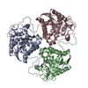
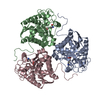
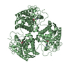
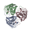
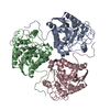
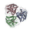
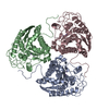
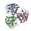
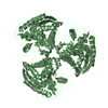
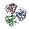
| Title | Amino Acid Sulfonamides as Transition-State Analogue Inhibitors of Arginase | ||||||
 Components Components | Arginase 1 | ||||||
 Keywords Keywords | HYDROLASE / arginase / sulfonamides / transition-state analogue | ||||||
| Function / homology |  Function and homology information Function and homology informationregulation of L-arginine import across plasma membrane / Urea cycle / collagen biosynthetic process / mammary gland involution / positive regulation of neutrophil mediated killing of fungus / negative regulation of T-helper 2 cell cytokine production / arginine metabolic process / arginase / arginase activity / urea cycle ...regulation of L-arginine import across plasma membrane / Urea cycle / collagen biosynthetic process / mammary gland involution / positive regulation of neutrophil mediated killing of fungus / negative regulation of T-helper 2 cell cytokine production / arginine metabolic process / arginase / arginase activity / urea cycle / response to selenium ion / response to methylmercury / response to nematode / response to manganese ion / response to steroid hormone / response to vitamin A / response to amine / response to herbicide / Neutrophil degranulation / response to zinc ion / response to vitamin E / defense response to protozoan / negative regulation of activated T cell proliferation / negative regulation of type II interferon-mediated signaling pathway / response to amino acid / maternal process involved in female pregnancy / cellular response to dexamethasone stimulus / response to axon injury / response to cadmium ion / cellular response to transforming growth factor beta stimulus / negative regulation of T cell proliferation / cellular response to interleukin-4 / positive regulation of endothelial cell proliferation / lung development / cellular response to glucagon stimulus / liver development / female pregnancy / response to peptide hormone / cellular response to hydrogen peroxide / manganese ion binding / cellular response to lipopolysaccharide / response to lipopolysaccharide / adaptive immune response / response to xenobiotic stimulus / innate immune response / neuronal cell body / : / identical protein binding / cytosol / cytoplasm Similarity search - Function | ||||||
| Biological species |  | ||||||
| Method |  X-RAY DIFFRACTION / SYNCHROTRON / MOLECULAR REPLACEMENT / Resolution: 2.8 Å X-RAY DIFFRACTION / SYNCHROTRON / MOLECULAR REPLACEMENT / Resolution: 2.8 Å | ||||||
 Authors Authors | Cama, E. / Shin, H. / Christianson, D.W. | ||||||
 Citation Citation | Journal: J.Am.Chem.Soc. / Year: 2003 Title: Design of Amino Acid Sulfonamides as Transition-State Analogue Inhibitors of Arginase Authors: Cama, E. / Shin, H. / Christianson, D.W. | ||||||
| History |
|
- Structure visualization
| Structure viewer | Molecule: MolmilJmol/JSmol |
|---|
- Downloads & links
-Download
| PDBx/mmCIF format | 1r1o.cif.gz | 188.8 KB | Display | PDBx/mmCIF format |
|---|---|---|---|---|
| PDB format | pdb1r1o.ent.gz | 151.1 KB | Display | PDB format |
| PDBx/mmJSON format | 1r1o.json.gz | Tree view | PDBx/mmJSON format | |
| Others |  Other downloads Other downloads |
-Validation report
| Arichive directory | https://data.pdbj.org/pub/pdb/validation_reports/r1/1r1oftp://data.pdbj.org/pub/pdb/validation_reports/r1/1r1o | HTTPS FTP |
|---|
-Related structure data
| Related structure data | |
|---|---|
| Similar structure data |


-Links
 PDBj
PDBj


- Assembly
| Deposited unit | 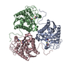
| ||||||||
|---|---|---|---|---|---|---|---|---|---|
| 1 |
| ||||||||
| Unit cell |
|
-Components
| #1: Protein | Mass: 35019.098 Da / Num. of mol.: 3 Source method: isolated from a genetically manipulated source Source: (gene. exp.)  #2: Chemical | ChemComp-MN /   Mass: 54.938 Da / Num. of mol.: 6 / Source method: obtained synthetically / Formula: Mn Mass: 54.938 Da / Num. of mol.: 6 / Source method: obtained synthetically / Formula: Mn#3: Chemical |   Mass: 228.290 Da / Num. of mol.: 3 / Source method: obtained synthetically / Formula: C5H12N2O4S2 Mass: 228.290 Da / Num. of mol.: 3 / Source method: obtained synthetically / Formula: C5H12N2O4S2#4: Water | ChemComp-HOH / |  Mass: 18.015 Da / Num. of mol.: 41 / Source method: isolated from a natural source / Formula: H2O Mass: 18.015 Da / Num. of mol.: 41 / Source method: isolated from a natural source / Formula: H2O |
|---|
-Experimental details
-Experiment
| Experiment | Method: X-RAY DIFFRACTION / Number of used crystals: 1 |
|---|
- Sample preparation
| Crystal | Density Matthews: 2.44 Å3/Da / Density % sol: 49.68 % | ||||||||||||||||||||||||||||||||||||||||||||||||||||||||
|---|---|---|---|---|---|---|---|---|---|---|---|---|---|---|---|---|---|---|---|---|---|---|---|---|---|---|---|---|---|---|---|---|---|---|---|---|---|---|---|---|---|---|---|---|---|---|---|---|---|---|---|---|---|---|---|---|---|
| Crystal grow | Temperature: 277 K / Method: vapor diffusion, hanging drop / pH: 8.5 Details: PEG 8000, bicine, manganese chloride, SDC, pH 8.5, VAPOR DIFFUSION, HANGING DROP, temperature 277K | ||||||||||||||||||||||||||||||||||||||||||||||||||||||||
| Crystal grow | *PLUS Temperature: 4 ℃ / Method: vapor diffusion, hanging drop | ||||||||||||||||||||||||||||||||||||||||||||||||||||||||
| Components of the solutions | *PLUS
|
-Data collection
| Diffraction | Mean temperature: 110 K |
|---|---|
| Diffraction source | Source: SYNCHROTRON / Site: CHESS  / Beamline: F1 / Wavelength: 0.916 Å / Beamline: F1 / Wavelength: 0.916 Å |
| Detector | Type: ADSC QUANTUM 4 / Detector: CCD / Date: Jan 17, 2002 |
| Radiation | Monochromator: Si(111) / Protocol: SINGLE WAVELENGTH / Monochromatic (M) / Laue (L): M / Scattering type: x-ray |
| Radiation wavelength | Wavelength: 0.916 Å / Relative weight: 1 |
| Reflection | Resolution: 2.8→30 Å / Num. obs: 23105 / % possible obs: 92.4 % / Observed criterion σ(F): 0 / Observed criterion σ(I): 0 |
| Reflection shell | Resolution: 2.8→2.9 Å / % possible all: 82.5 |
| Reflection | *PLUS Num. measured all: 204073 / Rmerge(I) obs: 0.098 |
| Reflection shell | *PLUS % possible obs: 82.5 % / Rmerge(I) obs: 0.287 |
- Processing
| Software |
| ||||||||||||||||||||||||||||||||||||
|---|---|---|---|---|---|---|---|---|---|---|---|---|---|---|---|---|---|---|---|---|---|---|---|---|---|---|---|---|---|---|---|---|---|---|---|---|---|
| Refinement | Method to determine structure: MOLECULAR REPLACEMENT / Resolution: 2.8→30 Å / Isotropic thermal model: RESTRAINED / Cross valid method: THROUGHOUT / σ(F): 0 / Stereochemistry target values: Engh & Huber
| ||||||||||||||||||||||||||||||||||||
| Refine analyze |
| ||||||||||||||||||||||||||||||||||||
| Refinement step | Cycle: LAST / Resolution: 2.8→30 Å
| ||||||||||||||||||||||||||||||||||||
| Refine LS restraints |
| ||||||||||||||||||||||||||||||||||||
| Refine LS restraints NCS | NCS model details: CONSTR | ||||||||||||||||||||||||||||||||||||
| Xplor file |
| ||||||||||||||||||||||||||||||||||||
| Software | *PLUS Name: CNS / Version: 1.1 / Classification: refinement | ||||||||||||||||||||||||||||||||||||
| Refinement | *PLUS % reflection Rfree: 5 % / Rfactor Rfree: 0.29 | ||||||||||||||||||||||||||||||||||||
| Solvent computation | *PLUS | ||||||||||||||||||||||||||||||||||||
| Displacement parameters | *PLUS | ||||||||||||||||||||||||||||||||||||
| Refine LS restraints | *PLUS
|
