 Movie
Movie Controller

[English] 日本語
 Yorodumi
- PDB-1qw9: Crystal structure of a family 51 alpha-L-arabinofuranosidase in c... -
+ Open data

- Basic information
| Entry | Database: PDB / ID: 1qw9 | |||||||||
|---|---|---|---|---|---|---|---|---|---|---|
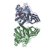
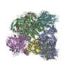
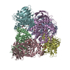
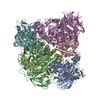
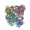
| Title | Crystal structure of a family 51 alpha-L-arabinofuranosidase in complex with 4-nitrophenyl-Ara | |||||||||
 Components Components | Alpha-L-arabinofuranosidase | |||||||||
 Keywords Keywords | HYDROLASE | |||||||||
| Function / homology |  Function and homology information Function and homology informationarabinan catabolic process / L-arabinose metabolic process / non-reducing end alpha-L-arabinofuranosidase / alpha-L-arabinofuranosidase activity / cytoplasm Similarity search - Function | |||||||||
| Biological species |   Geobacillus stearothermophilus (bacteria) Geobacillus stearothermophilus (bacteria) | |||||||||
| Method |  X-RAY DIFFRACTION / SYNCHROTRON / MOLECULAR REPLACEMENT / Resolution: 1.2 Å X-RAY DIFFRACTION / SYNCHROTRON / MOLECULAR REPLACEMENT / Resolution: 1.2 Å | |||||||||
 Authors Authors | Hoevel, K. / Shallom, D. / Niefind, K. / Belakhov, V. / Shoham, G. / Bassov, T. / Shoham, Y. / Schomburg, D. | |||||||||
 Citation Citation | Journal: Embo J. / Year: 2003 Title: Crystal structure and snapshots along the reaction pathway of a family 51 alpha-L-arabinofuranosidase Authors: Hoevel, K. / Shallom, D. / Niefind, K. / Belakhov, V. / Shoham, G. / Baasov, T. / Shoham, Y. / Schomburg, D. | |||||||||
| History |
|
- Structure visualization
| Structure viewer | Molecule: MolmilJmol/JSmol |
|---|
- Downloads & links
-Download
| PDBx/mmCIF format | 1qw9.cif.gz | 460.7 KB | Display | PDBx/mmCIF format |
|---|---|---|---|---|
| PDB format | pdb1qw9.ent.gz | 376.8 KB | Display | PDB format |
| PDBx/mmJSON format | 1qw9.json.gz | Tree view | PDBx/mmJSON format | |
| Others |  Other downloads Other downloads |
-Validation report
| Arichive directory | https://data.pdbj.org/pub/pdb/validation_reports/qw/1qw9ftp://data.pdbj.org/pub/pdb/validation_reports/qw/1qw9 | HTTPS FTP |
|---|
-Related structure data


-Links
 PDBj
PDBj- Assembly
| Deposited unit | 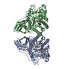
| |||||||||
|---|---|---|---|---|---|---|---|---|---|---|
| 1 | 
| |||||||||
| Unit cell |
| |||||||||
| Components on special symmetry positions |
|
-Components
| #1: Protein | Mass: 57226.863 Da / Num. of mol.: 2 / Mutation: E175A Source method: isolated from a genetically manipulated source Source: (gene. exp.) Geobacillus stearothermophilus (bacteria)Gene: ABFA / Plasmid: pET9d / Species (production host): Escherichia coli / Production host: References: UniProt: Q9XBQ3, non-reducing end alpha-L-arabinofuranosidase #2: Sugar |   Type: L-saccharide / Mass: 271.223 Da / Num. of mol.: 2 / Source method: obtained synthetically / Formula: C11H13NO7 Type: L-saccharide / Mass: 271.223 Da / Num. of mol.: 2 / Source method: obtained synthetically / Formula: C11H13NO7#3: Water | ChemComp-HOH / |  Mass: 18.015 Da / Num. of mol.: 1588 / Source method: isolated from a natural source / Formula: H2O Mass: 18.015 Da / Num. of mol.: 1588 / Source method: isolated from a natural source / Formula: H2O |
|---|
-Experimental details
-Experiment
| Experiment | Method: X-RAY DIFFRACTION / Number of used crystals: 1 |
|---|
- Sample preparation
| Crystal | Density Matthews: 2.71 Å3/Da / Density % sol: 54.66 % | |||||||||||||||||||||||||||||||||||
|---|---|---|---|---|---|---|---|---|---|---|---|---|---|---|---|---|---|---|---|---|---|---|---|---|---|---|---|---|---|---|---|---|---|---|---|---|
| Crystal grow | Temperature: 293 K / Method: vapor diffusion, sitting drop / pH: 8 Details: 18 % PEG 3350, 0.2 M NH4F, 5 % 2-propanol, 0.1 M Tris/HCl, pH 8.0, VAPOR DIFFUSION, SITTING DROP, temperature 293K | |||||||||||||||||||||||||||||||||||
| Crystal grow | *PLUS Method: vapor diffusion, sitting drop | |||||||||||||||||||||||||||||||||||
| Components of the solutions | *PLUS
|
-Data collection
| Diffraction | Mean temperature: 100 K |
|---|---|
| Diffraction source | Source: SYNCHROTRON / Site: EMBL/DESY, HAMBURG  / Beamline: BW7B / Wavelength: 0.8459 Å / Beamline: BW7B / Wavelength: 0.8459 Å |
| Detector | Type: MARRESEARCH / Detector: IMAGE PLATE / Date: Sep 16, 2002 |
| Radiation | Protocol: SINGLE WAVELENGTH / Monochromatic (M) / Laue (L): M / Scattering type: x-ray |
| Radiation wavelength | Wavelength: 0.8459 Å / Relative weight: 1 |
| Reflection | Resolution: 1.2→50 Å / Num. obs: 370234 / % possible obs: 99.1 % / Observed criterion σ(F): 0 / Observed criterion σ(I): 0 |
| Reflection shell | Resolution: 1.2→1.24 Å / % possible all: 97.9 |
| Reflection | *PLUS Num. obs: 370260 / Rmerge(I) obs: 0.049 |
| Reflection shell | *PLUS % possible obs: 97.9 % / Rmerge(I) obs: 0.383 / Mean I/σ(I) obs: 1.7 |
- Processing
| Software |
| ||||||||||||||||||||
|---|---|---|---|---|---|---|---|---|---|---|---|---|---|---|---|---|---|---|---|---|---|
| Refinement | Method to determine structure: MOLECULAR REPLACEMENT / Resolution: 1.2→50 Å / σ(F): 0 / Stereochemistry target values: Engh & Huber
| ||||||||||||||||||||
| Refinement step | Cycle: LAST / Resolution: 1.2→50 Å
| ||||||||||||||||||||
| Software | *PLUS Version: 5 / Classification: refinement | ||||||||||||||||||||
| Refinement | *PLUS Rfactor Rwork: 0.16 | ||||||||||||||||||||
| Solvent computation | *PLUS | ||||||||||||||||||||
| Displacement parameters | *PLUS | ||||||||||||||||||||
| Refine LS restraints | *PLUS
|
