 Movie
Movie Controller

[English] 日本語
 Yorodumi
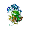
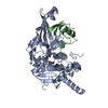
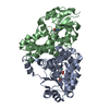
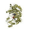
- PDB-1qq5: STRUCTURE OF L-2-HALOACID DEHALOGENASE FROM XANTHOBACTER AUTOTROPHICUS -
+ Open data

- Basic information
| Entry | Database: PDB / ID: 1qq5 | ||||||
|---|---|---|---|---|---|---|---|
| Title | STRUCTURE OF L-2-HALOACID DEHALOGENASE FROM XANTHOBACTER AUTOTROPHICUS | ||||||
 Components Components | PROTEIN (L-2-HALOACID DEHALOGENASE) | ||||||
 Keywords Keywords | HYDROLASE / L-2-HALOACID DEHALOGENASE | ||||||
| Function / homology |  Function and homology information Function and homology information | ||||||
| Biological species |  Xanthobacter autotrophicus (bacteria) Xanthobacter autotrophicus (bacteria) | ||||||
| Method |  X-RAY DIFFRACTION / SYNCHROTRON / MOLECULAR REPLACEMENT / Resolution: 1.52 Å X-RAY DIFFRACTION / SYNCHROTRON / MOLECULAR REPLACEMENT / Resolution: 1.52 Å | ||||||
 Authors Authors | Ridder, I.S. / Rozeboom, H.J. / Kalk, K.H. / Dijkstra, B.W. | ||||||
 Citation Citation | Journal: J.Biol.Chem. / Year: 1999 Title: Crystal structures of intermediates in the dehalogenation of haloalkanoates by L-2-haloacid dehalogenase. Authors: Ridder, I.S. / Rozeboom, H.J. / Kalk, K.H. / Dijkstra, B.W. #1: Journal: J.Biol.Chem. / Year: 1997Title: Three-Dimensional Structure of L-2-Haloacid Dehalogenase from Xanthobacter Autotrophicus Gj10 Complexed with the Substrate-Analogue Formate Authors: Ridder, I.S. / Rozeboom, H.J. / Kalk, K.H. / Janssen, D.B. / Dijkstra, B.W. #2: Journal: Protein Sci. / Year: 1995Title: Crystallization and Preliminary X-Ray Analysis of L-2-Haloacid Dehalogenase from Xanthobacter Autotrophicus Gj10 Authors: Ridder, I.S. / Rozeboom, H.J. / Kingma, J. / Janssen, D.B. / Dijkstra, B.W. | ||||||
| History |
|
- Structure visualization
| Structure viewer | Molecule: MolmilJmol/JSmol |
|---|
- Downloads & links
-Download
| PDBx/mmCIF format | 1qq5.cif.gz | 122.4 KB | Display | PDBx/mmCIF format |
|---|---|---|---|---|
| PDB format | pdb1qq5.ent.gz | 94.6 KB | Display | PDB format |
| PDBx/mmJSON format | 1qq5.json.gz | Tree view | PDBx/mmJSON format | |
| Others |  Other downloads Other downloads |
-Validation report
| Arichive directory | https://data.pdbj.org/pub/pdb/validation_reports/qq/1qq5ftp://data.pdbj.org/pub/pdb/validation_reports/qq/1qq5 | HTTPS FTP |
|---|
-Related structure data
| Related structure data |  1qq6C  1qq7C 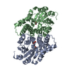 1aq6S C: citing same article ( S: Starting model for refinement |
|---|---|
| Similar structure data |


-Links
 PDBj
PDBj

- Assembly
| Deposited unit | 
| ||||||||
|---|---|---|---|---|---|---|---|---|---|
| 1 |
| ||||||||
| Unit cell |
|
-Components
| #1: Protein | Mass: 27524.398 Da / Num. of mol.: 2 / Source method: isolated from a natural source Details: SUBSTRATE ANALOGUE FORMATE PRESENT IN BOTH ACTIVE SITES Source: (natural) Xanthobacter autotrophicus (bacteria) / Strain: GJ10 / References: UniProt: Q60099, (S)-2-haloacid dehalogenase#2: Chemical |   Mass: 46.025 Da / Num. of mol.: 2 / Source method: obtained synthetically / Formula: CH2O2 Mass: 46.025 Da / Num. of mol.: 2 / Source method: obtained synthetically / Formula: CH2O2#3: Water | ChemComp-HOH / |  Mass: 18.015 Da / Num. of mol.: 565 / Source method: isolated from a natural source / Formula: H2O Mass: 18.015 Da / Num. of mol.: 565 / Source method: isolated from a natural source / Formula: H2O |
|---|
-Experimental details
-Experiment
| Experiment | Method: X-RAY DIFFRACTION / Number of used crystals: 1 |
|---|
- Sample preparation
| Crystal | Density Matthews: 2 Å3/Da / Density % sol: 38 % | ||||||||||||||||||||||||||||||||||||||||
|---|---|---|---|---|---|---|---|---|---|---|---|---|---|---|---|---|---|---|---|---|---|---|---|---|---|---|---|---|---|---|---|---|---|---|---|---|---|---|---|---|---|
| Crystal grow | pH: 7 / Details: pH 7.00 | ||||||||||||||||||||||||||||||||||||||||
| Crystal | *PLUS | ||||||||||||||||||||||||||||||||||||||||
| Crystal grow | *PLUS Method: vapor diffusion, hanging dropDetails: used to seeding, Ridder, I.S., (1995) Protein Sci., 4, 2619. PH range low: 7 / PH range high: 6.8 | ||||||||||||||||||||||||||||||||||||||||
| Components of the solutions | *PLUS
|
-Data collection
| Diffraction | Mean temperature: 100 K |
|---|---|
| Diffraction source | Source: SYNCHROTRON / Site: ESRF  / Beamline: ID14-3 / Wavelength: 0.9475 / Beamline: ID14-3 / Wavelength: 0.9475 |
| Detector | Type: MARRESEARCH / Detector: CCD / Date: Apr 1, 1998 |
| Radiation | Protocol: SINGLE WAVELENGTH / Monochromatic (M) / Laue (L): M / Scattering type: x-ray |
| Radiation wavelength | Wavelength: 0.9475 Å / Relative weight: 1 |
| Reflection | Resolution: 1.52→30 Å / Num. obs: 63057 / % possible obs: 92.7 % / Observed criterion σ(I): -3 / Redundancy: 4.9 % / Biso Wilson estimate: 19.4 Å2 / Rmerge(I) obs: 0.064 / Net I/σ(I): 24.7 |
| Reflection shell | Resolution: 1.52→1.55 Å / Redundancy: 2.9 % / Rmerge(I) obs: 0.351 / % possible all: 81.4 |
| Reflection | *PLUS Highest resolution: 1.52 Å / Lowest resolution: 30 Å / Observed criterion σ(I): -3 / Redundancy: 4.9 % / Num. measured all: 306585 / Biso Wilson estimate: 19.4 Å2 |
| Reflection shell | *PLUS % possible obs: 81.4 % / Mean I/σ(I) obs: 2.2 |
- Processing
| Software |
| ||||||||||||||||||||||||||||||||||||||||||||||||||||||||||||||||||||||||||||||||
|---|---|---|---|---|---|---|---|---|---|---|---|---|---|---|---|---|---|---|---|---|---|---|---|---|---|---|---|---|---|---|---|---|---|---|---|---|---|---|---|---|---|---|---|---|---|---|---|---|---|---|---|---|---|---|---|---|---|---|---|---|---|---|---|---|---|---|---|---|---|---|---|---|---|---|---|---|---|---|---|---|---|
| Refinement | Method to determine structure: MOLECULAR REPLACEMENT Starting model: 1AQ6 Resolution: 1.52→20 Å / Rfactor Rfree error: 0.004 / Data cutoff high rms absF: 2391656.32 / Isotropic thermal model: RESTRAINED / Cross valid method: THROUGHOUT / σ(F): 0 / Stereochemistry target values: ENGH & HUBER Details: WEIGTH ON OMEGA DIHEDRAL ANGLE SET TO 20% OF VALUE IN CNS FORCE FIELD
| ||||||||||||||||||||||||||||||||||||||||||||||||||||||||||||||||||||||||||||||||
| Solvent computation | Solvent model: FLAT MODEL / Bsol: 61.22 Å2 / ksol: 0.36 e/Å3 | ||||||||||||||||||||||||||||||||||||||||||||||||||||||||||||||||||||||||||||||||
| Displacement parameters | Biso mean: 25.4 Å2
| ||||||||||||||||||||||||||||||||||||||||||||||||||||||||||||||||||||||||||||||||
| Refine analyze |
| ||||||||||||||||||||||||||||||||||||||||||||||||||||||||||||||||||||||||||||||||
| Refinement step | Cycle: LAST / Resolution: 1.52→20 Å
| ||||||||||||||||||||||||||||||||||||||||||||||||||||||||||||||||||||||||||||||||
| Refine LS restraints |
| ||||||||||||||||||||||||||||||||||||||||||||||||||||||||||||||||||||||||||||||||
| LS refinement shell | Resolution: 1.52→1.62 Å / Rfactor Rfree error: 0.01 / Total num. of bins used: 6
| ||||||||||||||||||||||||||||||||||||||||||||||||||||||||||||||||||||||||||||||||
| Xplor file |
| ||||||||||||||||||||||||||||||||||||||||||||||||||||||||||||||||||||||||||||||||
| Software | *PLUS Name: CNS / Version: 0.5 / Classification: refinement | ||||||||||||||||||||||||||||||||||||||||||||||||||||||||||||||||||||||||||||||||
| Refinement | *PLUS Lowest resolution: 20 Å / Rfactor obs: 0.198 | ||||||||||||||||||||||||||||||||||||||||||||||||||||||||||||||||||||||||||||||||
| Solvent computation | *PLUS | ||||||||||||||||||||||||||||||||||||||||||||||||||||||||||||||||||||||||||||||||
| Displacement parameters | *PLUS | ||||||||||||||||||||||||||||||||||||||||||||||||||||||||||||||||||||||||||||||||
| Refine LS restraints | *PLUS
| ||||||||||||||||||||||||||||||||||||||||||||||||||||||||||||||||||||||||||||||||
| LS refinement shell | *PLUS Rfactor Rfree: 0.301 |
