 Movie
Movie Controller

[English] 日本語
 Yorodumi
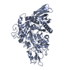
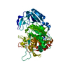
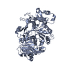
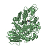
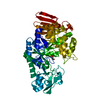
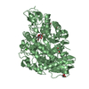
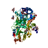
- PDB-3vnq: Co-crystal structure of NRPS adenylation protein CytC1 with ATP f... -
+ Open data

- Basic information
| Entry | Database: PDB / ID: 3vnq | ||||||
|---|---|---|---|---|---|---|---|
| Title | Co-crystal structure of NRPS adenylation protein CytC1 with ATP from streptomyces | ||||||
 Components Components | NRPS adenylation protein CytC1 | ||||||
 Keywords Keywords | LIGASE / Adenylation / ATP-binding / Non-ribosomal peptide synthese / NRPS / Streptomyces | ||||||
| Function / homology | ANL, N-terminal domain / ANL, C-terminal domain / GMP Synthetase; Chain A, domain 3 / Rossmann fold / 2-Layer Sandwich / 3-Layer(aba) Sandwich / Alpha Beta / ADENOSINE-5'-TRIPHOSPHATE Function and homology information Function and homology information | ||||||
| Biological species |  Streptomyces sp. (bacteria) Streptomyces sp. (bacteria) | ||||||
| Method |  X-RAY DIFFRACTION / SYNCHROTRON / MOLECULAR REPLACEMENT / Resolution: 2.1 Å X-RAY DIFFRACTION / SYNCHROTRON / MOLECULAR REPLACEMENT / Resolution: 2.1 Å | ||||||
 Authors Authors | Okumura, H. / Ueki, M. / Shiro, Y. / Osada, H. | ||||||
 Citation Citation | Journal: To be Published Title: Substrate recognition mechanism of NRPS adenylation protein from Streptomyces Authors: Ueki, M. / Okumura, H. / Osada, H. | ||||||
| History |
|
- Structure visualization
| Structure viewer | Molecule: MolmilJmol/JSmol |
|---|
- Downloads & links
-Download
| PDBx/mmCIF format | 3vnq.cif.gz | 116.8 KB | Display | PDBx/mmCIF format |
|---|---|---|---|---|
| PDB format | pdb3vnq.ent.gz | 87.7 KB | Display | PDB format |
| PDBx/mmJSON format | 3vnq.json.gz | Tree view | PDBx/mmJSON format | |
| Others |  Other downloads Other downloads |
-Validation report
| Arichive directory | https://data.pdbj.org/pub/pdb/validation_reports/vn/3vnqftp://data.pdbj.org/pub/pdb/validation_reports/vn/3vnq | HTTPS FTP |
|---|
-Related structure data
| Related structure data |  3vnrC  3vnsC 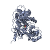 1amuS C: citing same article ( S: Starting model for refinement |
|---|---|
| Similar structure data |


-Links
 PDBj
PDBj- Assembly
| Deposited unit | 
| |||||||||
|---|---|---|---|---|---|---|---|---|---|---|
| 1 |
| |||||||||
| Unit cell |
| |||||||||
| Components on special symmetry positions |
|
-Components
| #1: Protein | Mass: 58992.520 Da / Num. of mol.: 1 Source method: isolated from a genetically manipulated source Source: (gene. exp.) Streptomyces sp. (bacteria) / Strain: RK95-74 / Production host: |
|---|---|
| #2: Chemical | ChemComp-ATP /   Mass: 507.181 Da / Num. of mol.: 1 / Source method: obtained synthetically / Formula: C10H16N5O13P3 / Comment: ATP, energy-carrying molecule*YM Mass: 507.181 Da / Num. of mol.: 1 / Source method: obtained synthetically / Formula: C10H16N5O13P3 / Comment: ATP, energy-carrying molecule*YM |
| #3: Water | ChemComp-HOH /  Mass: 18.015 Da / Num. of mol.: 281 / Source method: isolated from a natural source / Formula: H2O Mass: 18.015 Da / Num. of mol.: 281 / Source method: isolated from a natural source / Formula: H2O |
| Sequence details | A SEQUENCE DATABASE REFERENCE FOR THIS PROTEIN DOES NOT CURRENTLY EXIST. |
-Experimental details
-Experiment
| Experiment | Method: X-RAY DIFFRACTION / Number of used crystals: 1 |
|---|
- Sample preparation
| Crystal | Density Matthews: 2.69 Å3/Da / Density % sol: 54.26 % |
|---|---|
| Crystal grow | Temperature: 293 K / Method: vapor diffusion, sitting drop / pH: 7.2 Details: 0.1M ammonium sulfate, 24-32% PEG5000 monomethyl ether, 1mM ATP, 100mM Hepes buffer, pH 7.2, VAPOR DIFFUSION, SITTING DROP, temperature 293K |
-Data collection
| Diffraction | Mean temperature: 100 K |
|---|---|
| Diffraction source | Source: SYNCHROTRON / Site: SPring-8  / Beamline: BL44B2 / Wavelength: 1 Å / Beamline: BL44B2 / Wavelength: 1 Å |
| Detector | Type: ADSC QUANTUM 210 / Detector: CCD / Date: Dec 14, 2006 / Details: mirrors |
| Radiation | Monochromator: Si(111) / Protocol: SINGLE WAVELENGTH / Monochromatic (M) / Laue (L): M / Scattering type: x-ray |
| Radiation wavelength | Wavelength: 1 Å / Relative weight: 1 |
| Reflection | Resolution: 2.1→50 Å / Num. obs: 37003 / % possible obs: 100 % / Observed criterion σ(F): 0 / Observed criterion σ(I): -3 / Redundancy: 15.1 % / Biso Wilson estimate: 27.52 Å2 / Rmerge(I) obs: 0.066 / Rsym value: 0.066 / Net I/σ(I): 53.384 |
| Reflection shell | Resolution: 2.1→2.14 Å / Redundancy: 15.1 % / Rmerge(I) obs: 0.405 / Mean I/σ(I) obs: 7.878 / Rsym value: 0.405 / % possible all: 100 |
- Processing
| Software |
| ||||||||||||||||||||||||||||||||||||||||||||||||||||||||||||||||||||||||||||||||||||||||||||||||||
|---|---|---|---|---|---|---|---|---|---|---|---|---|---|---|---|---|---|---|---|---|---|---|---|---|---|---|---|---|---|---|---|---|---|---|---|---|---|---|---|---|---|---|---|---|---|---|---|---|---|---|---|---|---|---|---|---|---|---|---|---|---|---|---|---|---|---|---|---|---|---|---|---|---|---|---|---|---|---|---|---|---|---|---|---|---|---|---|---|---|---|---|---|---|---|---|---|---|---|---|
| Refinement | Method to determine structure: MOLECULAR REPLACEMENT Starting model: 1AMU Resolution: 2.1→34.915 Å / Occupancy max: 1 / Occupancy min: 0.5 / FOM work R set: 0.8579 / SU ML: 0.26 / σ(F): 0 / Phase error: 21.27 / Stereochemistry target values: ML
| ||||||||||||||||||||||||||||||||||||||||||||||||||||||||||||||||||||||||||||||||||||||||||||||||||
| Solvent computation | Shrinkage radii: 0.9 Å / VDW probe radii: 1.11 Å / Solvent model: FLAT BULK SOLVENT MODEL / Bsol: 40.447 Å2 / ksol: 0.351 e/Å3 | ||||||||||||||||||||||||||||||||||||||||||||||||||||||||||||||||||||||||||||||||||||||||||||||||||
| Displacement parameters | Biso max: 89.75 Å2 / Biso mean: 31.1802 Å2 / Biso min: 11.18 Å2
| ||||||||||||||||||||||||||||||||||||||||||||||||||||||||||||||||||||||||||||||||||||||||||||||||||
| Refinement step | Cycle: LAST / Resolution: 2.1→34.915 Å
| ||||||||||||||||||||||||||||||||||||||||||||||||||||||||||||||||||||||||||||||||||||||||||||||||||
| Refine LS restraints |
| ||||||||||||||||||||||||||||||||||||||||||||||||||||||||||||||||||||||||||||||||||||||||||||||||||
| LS refinement shell | Refine-ID: X-RAY DIFFRACTION / Total num. of bins used: 13
|
