 Movie
Movie Controller


+ Open data

- Basic information
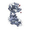
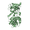
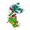
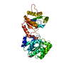
| Entry | Database: PDB / ID: 1qpg | ||||||
|---|---|---|---|---|---|---|---|
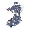
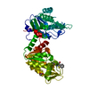
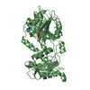
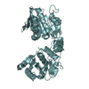
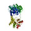
| Title | 3-PHOSPHOGLYCERATE KINASE, MUTATION R65Q | ||||||
 Components Components | 3-PHOSPHOGLYCERATE KINASE | ||||||
 Keywords Keywords | PHOSPHOTRANSFERASE (CARBOXYL ACCEPTOR) / KINASE / ACETYLATION / GLYCOLYSIS | ||||||
| Function / homology |  Function and homology information Function and homology informationGluconeogenesis / Glycolysis / phosphoglycerate kinase / phosphoglycerate kinase activity / glycolytic process / gluconeogenesis / ADP binding / mitochondrion / ATP binding / metal ion binding ...Gluconeogenesis / Glycolysis / phosphoglycerate kinase / phosphoglycerate kinase activity / glycolytic process / gluconeogenesis / ADP binding / mitochondrion / ATP binding / metal ion binding / plasma membrane / cytoplasm / cytosol Similarity search - Function | ||||||
| Biological species |  | ||||||
| Method |  X-RAY DIFFRACTION / Resolution: 2.4 Å X-RAY DIFFRACTION / Resolution: 2.4 Å | ||||||
 Authors Authors | Mcphillips, T.M. / Hsu, B.T. / Sherman, M.A. / Mas, M.T. / Rees, D.C. | ||||||
 Citation Citation | Journal: Biochemistry / Year: 1996 Title: Structure of the R65Q mutant of yeast 3-phosphoglycerate kinase complexed with Mg-AMP-PNP and 3-phospho-D-glycerate. Authors: McPhillips, T.M. / Hsu, B.T. / Sherman, M.A. / Mas, M.T. / Rees, D.C. #1: Journal: Protein Sci. / Year: 1992Title: Characterization of the Structure and Properties of the His 62-->Ala and Arg 38-->Ala Mutants of Yeast Phosphoglycerate Kinase: An Investigation of the Catalytic and Activatory Sites by Site- ...Title: Characterization of the Structure and Properties of the His 62-->Ala and Arg 38-->Ala Mutants of Yeast Phosphoglycerate Kinase: An Investigation of the Catalytic and Activatory Sites by Site-Directed Mutagenesis and NMR Authors: Sherman, M.A. / Fairbrother, W.J. / Mas, M.T. #2: Journal: Protein Eng. / Year: 1991Title: Site-Directed Mutations of Arginine 65 at the Periphery of the Active Site Cleft of Yeast 3-Phosphoglycerate Kinase Enhance the Catalytic Activity and Eliminate Anion-Dependent Activation Authors: Sherman, M.A. / Dean, S.A. / Mathiowetz, A.M. / Mas, M.T. #3: Journal: J.Biol.Chem. / Year: 1990Title: Probing the Role of Arginines and Histidines in the Catalytic Function and Activation of Yeast 3-Phosphoglycerate Kinase by Site-Directed Mutagenesis Authors: Sherman, M.A. / Szpikowska, B.K. / Dean, S.A. / Mathiowetz, A.M. / Mcqueen, N.L. / Mas, M.T. | ||||||
| History |
|
- Structure visualization
| Structure viewer | Molecule: MolmilJmol/JSmol |
|---|
- Downloads & links
-Download
| PDBx/mmCIF format | 1qpg.cif.gz | 94.2 KB | Display | PDBx/mmCIF format |
|---|---|---|---|---|
| PDB format | pdb1qpg.ent.gz | 70.5 KB | Display | PDB format |
| PDBx/mmJSON format | 1qpg.json.gz | Tree view | PDBx/mmJSON format | |
| Others |  Other downloads Other downloads |
-Validation report
| Arichive directory | https://data.pdbj.org/pub/pdb/validation_reports/qp/1qpgftp://data.pdbj.org/pub/pdb/validation_reports/qp/1qpg | HTTPS FTP |
|---|
-Related structure data
| Similar structure data |
|---|

-Links
 PDBj
PDBj
- Assembly
| Deposited unit | 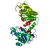
| ||||||||
|---|---|---|---|---|---|---|---|---|---|
| 1 |
| ||||||||
| Unit cell |
|
-Components
| #1: Protein | Mass: 44641.059 Da / Num. of mol.: 1 / Mutation: R65Q Source method: isolated from a genetically manipulated source Source: (gene. exp.) Plasmid: YEP9T-PGK / Production host: |
|---|---|
| #2: Chemical | ChemComp-MAP /   Mass: 529.493 Da / Num. of mol.: 1 / Source method: obtained synthetically / Formula: C10H16MgN6O12P3 Mass: 529.493 Da / Num. of mol.: 1 / Source method: obtained synthetically / Formula: C10H16MgN6O12P3 |
| #3: Chemical | ChemComp-3PG /   Mass: 186.057 Da / Num. of mol.: 1 / Source method: obtained synthetically / Formula: C3H7O7P Mass: 186.057 Da / Num. of mol.: 1 / Source method: obtained synthetically / Formula: C3H7O7P |
| #4: Water | ChemComp-HOH /  Mass: 18.015 Da / Num. of mol.: 82 / Source method: isolated from a natural source / Formula: H2O Mass: 18.015 Da / Num. of mol.: 82 / Source method: isolated from a natural source / Formula: H2O |
-Experimental details
-Experiment
| Experiment | Method: X-RAY DIFFRACTION |
|---|
- Sample preparation
| Crystal | Density Matthews: 2.62 Å3/Da / Density % sol: 53.12 % | ||||||||||||||||||||||||||||||||||||||||||||||||||||||||||||
|---|---|---|---|---|---|---|---|---|---|---|---|---|---|---|---|---|---|---|---|---|---|---|---|---|---|---|---|---|---|---|---|---|---|---|---|---|---|---|---|---|---|---|---|---|---|---|---|---|---|---|---|---|---|---|---|---|---|---|---|---|---|
| Crystal grow | *PLUS pH: 7.5 / Method: vapor diffusion, hanging drop | ||||||||||||||||||||||||||||||||||||||||||||||||||||||||||||
| Components of the solutions | *PLUS
|
-Data collection
| Diffraction source | Wavelength: 1.5418 |
|---|---|
| Detector | Type: SIEMENS / Detector: AREA DETECTOR / Date: Sep 30, 1991 |
| Radiation | Monochromatic (M) / Laue (L): M / Scattering type: x-ray |
| Radiation wavelength | Wavelength: 1.5418 Å / Relative weight: 1 |
| Reflection | Resolution: 2.43→40.7 Å / Num. obs: 16281 / % possible obs: 92.4 % / Observed criterion σ(I): 0 / Redundancy: 6.2 % / Rmerge(I) obs: 0.076 |
| Reflection | *PLUS Num. measured all: 100828 |
- Processing
| Software |
| ||||||||||||||||||||||||||||||||||||||||||||||||||||||||||||
|---|---|---|---|---|---|---|---|---|---|---|---|---|---|---|---|---|---|---|---|---|---|---|---|---|---|---|---|---|---|---|---|---|---|---|---|---|---|---|---|---|---|---|---|---|---|---|---|---|---|---|---|---|---|---|---|---|---|---|---|---|---|
| Refinement | Resolution: 2.4→8 Å / σ(F): 0
| ||||||||||||||||||||||||||||||||||||||||||||||||||||||||||||
| Displacement parameters | Biso mean: 33.9 Å2 | ||||||||||||||||||||||||||||||||||||||||||||||||||||||||||||
| Refinement step | Cycle: LAST / Resolution: 2.4→8 Å
| ||||||||||||||||||||||||||||||||||||||||||||||||||||||||||||
| Refine LS restraints |
| ||||||||||||||||||||||||||||||||||||||||||||||||||||||||||||
| Software | *PLUS Name: X-PLOR / Classification: refinement | ||||||||||||||||||||||||||||||||||||||||||||||||||||||||||||
| Refinement | *PLUS | ||||||||||||||||||||||||||||||||||||||||||||||||||||||||||||
| Solvent computation | *PLUS | ||||||||||||||||||||||||||||||||||||||||||||||||||||||||||||
| Displacement parameters | *PLUS | ||||||||||||||||||||||||||||||||||||||||||||||||||||||||||||
| Refine LS restraints | *PLUS
|
