 Movie
Movie Controller


+ Open data

- Basic information
| Entry | Database: PDB / ID: 1qlw | ||||||
|---|---|---|---|---|---|---|---|
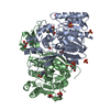
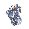
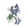
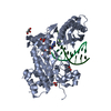
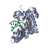
| Title | The Atomic Resolution Structure of a Novel Bacterial Esterase | ||||||
 Components Components | ESTERASE | ||||||
 Keywords Keywords | HYDROLASE(CARBOXYLIC ESTERASE) / ESTERASE / ANISOTROPIC REFINEMENT / ATOMIC RESOLUTION / ALPHA/BETA HYDROLASE | ||||||
| Function / homology | : / Alpha/Beta hydrolase fold, catalytic domain / Alpha/Beta hydrolase fold / Rossmann fold / 3-Layer(aba) Sandwich / Alpha Beta / Carboxylesterase Function and homology information Function and homology information | ||||||
| Biological species |  ALCALIGENES SP. (bacteria) ALCALIGENES SP. (bacteria) | ||||||
| Method |  X-RAY DIFFRACTION / SYNCHROTRON / MIR / Resolution: 1.09 Å X-RAY DIFFRACTION / SYNCHROTRON / MIR / Resolution: 1.09 Å | ||||||
 Authors Authors | Bourne, P.C. / Isupov, M.N. / Littlechild, J.A. | ||||||
 Citation Citation | Journal: Structure / Year: 2000 Title: The Atomic Resolution Structure of a Novel Bacterial Esterase Authors: Bourne, P.C. / Isupov, M.N. / Littlechild, J.A. #1: Journal: Acta Crystallogr.,Sect.D / Year: 1999 Title: Crystallization and Preliminary X-Ray Diffraction Studies of a Novel Bacterial Esterase Authors: Bourne, P.C. / Isupov, M.N. / Littlechild, J.A. | ||||||
| History |
|
- Structure visualization
| Structure viewer | Molecule: MolmilJmol/JSmol |
|---|
- Downloads & links
-Download
| PDBx/mmCIF format | 1qlw.cif.gz | 277 KB | Display | PDBx/mmCIF format |
|---|---|---|---|---|
| PDB format | pdb1qlw.ent.gz | 225.7 KB | Display | PDB format |
| PDBx/mmJSON format | 1qlw.json.gz | Tree view | PDBx/mmJSON format | |
| Others |  Other downloads Other downloads |
-Validation report
| Arichive directory | https://data.pdbj.org/pub/pdb/validation_reports/ql/1qlwftp://data.pdbj.org/pub/pdb/validation_reports/ql/1qlw | HTTPS FTP |
|---|
-Related structure data


-Links
 PDBj
PDBj- Assembly
| Deposited unit | 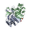
| ||||||||
|---|---|---|---|---|---|---|---|---|---|
| 1 |
| ||||||||
| Unit cell |
| ||||||||
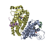
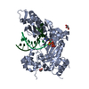
| Noncrystallographic symmetry (NCS) | NCS oper: (Code: given Matrix: (-0.99234, -0.02182, 0.12158), Vector: Details | BIOLOGICAL_UNIT: DIMERIC | |
-Components
| #1: Protein | Mass: 35686.770 Da / Num. of mol.: 2 Source method: isolated from a genetically manipulated source Details: ALPHA/BETA HYDROLASE FOLD / Source: (gene. exp.) ALCALIGENES SP. (bacteria) / Production host: AGROBACTERIUM SP. (bacteria) / References: UniProt: Q7SIA5*PLUS#2: Chemical |   Mass: 96.063 Da / Num. of mol.: 3 / Source method: obtained synthetically / Formula: SO4 Mass: 96.063 Da / Num. of mol.: 3 / Source method: obtained synthetically / Formula: SO4#3: Water | ChemComp-HOH / |  Mass: 18.015 Da / Num. of mol.: 715 / Source method: isolated from a natural source / Formula: H2O Mass: 18.015 Da / Num. of mol.: 715 / Source method: isolated from a natural source / Formula: H2OHas protein modification | Y | |
|---|
-Experimental details
-Experiment
| Experiment | Method: X-RAY DIFFRACTION / Number of used crystals: 1 |
|---|
- Sample preparation
| Crystal | Density Matthews: 2.37 Å3/Da / Density % sol: 48.03 % | ||||||||||||||||||||||||||||||
|---|---|---|---|---|---|---|---|---|---|---|---|---|---|---|---|---|---|---|---|---|---|---|---|---|---|---|---|---|---|---|---|
| Crystal grow | pH: 7 / Details: PH 4.6 | ||||||||||||||||||||||||||||||
| Crystal grow | *PLUS Method: vapor diffusion, hanging dropDetails: Bourne, P.C., (1999) Acta Crystallogr.,Sect.D, 55, 915. | ||||||||||||||||||||||||||||||
| Components of the solutions | *PLUS
|
-Data collection
| Diffraction | Mean temperature: 100 K |
|---|---|
| Diffraction source | Source: SYNCHROTRON / Site: EMBL/DESY, HAMBURG  / Beamline: BW7B / Wavelength: 0.8373 / Beamline: BW7B / Wavelength: 0.8373 |
| Detector | Type: MARRESEARCH / Detector: IMAGE PLATE / Date: Nov 15, 1997 / Details: BENT MIRROR |
| Radiation | Monochromator: HAMBURG / Protocol: SINGLE WAVELENGTH / Monochromatic (M) / Laue (L): M / Scattering type: x-ray |
| Radiation wavelength | Wavelength: 0.8373 Å / Relative weight: 1 |
| Reflection | Resolution: 1.09→28 Å / Num. obs: 274290 / % possible obs: 99.4 % / Observed criterion σ(I): 0 / Redundancy: 3.8 % / Biso Wilson estimate: 14.5 Å2 / Rmerge(I) obs: 0.094 / Net I/σ(I): 14.5 |
| Reflection shell | Resolution: 1.09→1.12 Å / Redundancy: 3.6 % / Rmerge(I) obs: 0.36 / Mean I/σ(I) obs: 2.3 / % possible all: 98.2 |
| Reflection shell | *PLUS % possible obs: 98.2 % |
- Processing
| Software |
| ||||||||||||||||||||||||||||||||||||||||||||||||||||||||||||||||||||||||||||||||||||
|---|---|---|---|---|---|---|---|---|---|---|---|---|---|---|---|---|---|---|---|---|---|---|---|---|---|---|---|---|---|---|---|---|---|---|---|---|---|---|---|---|---|---|---|---|---|---|---|---|---|---|---|---|---|---|---|---|---|---|---|---|---|---|---|---|---|---|---|---|---|---|---|---|---|---|---|---|---|---|---|---|---|---|---|---|---|
| Refinement | Method to determine structure: MIR / Resolution: 1.09→30 Å / SU B: 0.296 / SU ML: 0.015 / Cross valid method: THROUGHOUT / σ(F): 0 / ESU R: 0.025 / ESU R Free: 0.025 Details: THE C-TERMINAL RESIDUE WAS NOT SEEN IN THE DENSITY MAPS
| ||||||||||||||||||||||||||||||||||||||||||||||||||||||||||||||||||||||||||||||||||||
| Displacement parameters | Biso mean: 21.4 Å2
| ||||||||||||||||||||||||||||||||||||||||||||||||||||||||||||||||||||||||||||||||||||
| Refinement step | Cycle: LAST / Resolution: 1.09→30 Å
| ||||||||||||||||||||||||||||||||||||||||||||||||||||||||||||||||||||||||||||||||||||
| Refine LS restraints |
| ||||||||||||||||||||||||||||||||||||||||||||||||||||||||||||||||||||||||||||||||||||
| Software | *PLUS Name: REFMAC / Classification: refinement | ||||||||||||||||||||||||||||||||||||||||||||||||||||||||||||||||||||||||||||||||||||
| Refinement | *PLUS Rfactor obs: 0.1429 / % reflection Rfree: 5 % | ||||||||||||||||||||||||||||||||||||||||||||||||||||||||||||||||||||||||||||||||||||
| Solvent computation | *PLUS | ||||||||||||||||||||||||||||||||||||||||||||||||||||||||||||||||||||||||||||||||||||
| Displacement parameters | *PLUS |
