 Movie
Movie Controller


+ Open data

- Basic information
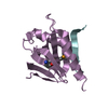
| Entry | Database: PDB / ID: 1qk8 | ||||||
|---|---|---|---|---|---|---|---|
| Title | TRYPAREDOXIN-I FROM CRITHIDIA FASCICULATA | ||||||
 Components Components | TRYPAREDOXIN-I | ||||||
 Keywords Keywords | TRYPAREDOXIN / CRITHIDIA FASCICULATA / THIOREDOXIN / TRYPANOSOME / ANOMALOUS DISPERSION / OXIDATIVE STRESS / OXIDOREDUCTASE | ||||||
| Function / homology |  Function and homology information Function and homology informationthioredoxin-disulfide reductase (NADPH) activity / negative regulation of Wnt signaling pathway / negative regulation of protein ubiquitination / nucleus Similarity search - Function | ||||||
| Biological species |  CRITHIDIA FASCICULATA (eukaryote) CRITHIDIA FASCICULATA (eukaryote) | ||||||
| Method |  X-RAY DIFFRACTION / SYNCHROTRON / MAD / Resolution: 1.4 Å X-RAY DIFFRACTION / SYNCHROTRON / MAD / Resolution: 1.4 Å | ||||||
 Authors Authors | Alphey, M.S. / Hunter, W.N. | ||||||
 Citation Citation | Journal: J.Biol.Chem. / Year: 1999 Title: The High Resolution Crystal Structure of Recombinant Crithidia Fasciculata Tryparedoxin-I Authors: Alphey, M.S. / Leonard, G.A. / Gourley, D.G. / Tetaud, E. / Fairlamb, A.H. / Hunter, W.N. | ||||||
| History |
| ||||||
| Remark 650 | HELIX DETERMINATION METHOD: DSSP | ||||||
| Remark 700 | SHEET DETERMINATION METHOD: DSSP |
- Structure visualization
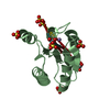
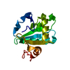
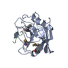
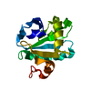
| Structure viewer | Molecule: MolmilJmol/JSmol |
|---|
- Downloads & links
-Download
| PDBx/mmCIF format | 1qk8.cif.gz | 46.3 KB | Display | PDBx/mmCIF format |
|---|---|---|---|---|
| PDB format | pdb1qk8.ent.gz | 32.3 KB | Display | PDB format |
| PDBx/mmJSON format | 1qk8.json.gz | Tree view | PDBx/mmJSON format | |
| Others |  Other downloads Other downloads |
-Validation report
| Summary document | 1qk8_validation.pdf.gz | 414 KB | Display | wwPDB validaton report |
|---|---|---|---|---|
| Full document | 1qk8_full_validation.pdf.gz | 414.8 KB | Display | |
| Data in XML | 1qk8_validation.xml.gz | 11.9 KB | Display | |
| Data in CIF | 1qk8_validation.cif.gz | 16.7 KB | Display | |
| Arichive directory | https://data.pdbj.org/pub/pdb/validation_reports/qk/1qk8ftp://data.pdbj.org/pub/pdb/validation_reports/qk/1qk8 | HTTPS FTP |
-Related structure data
| Similar structure data |
|---|


-Links
 PDBj
PDBj- Assembly
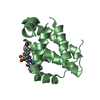
| Deposited unit | 
| ||||||||
|---|---|---|---|---|---|---|---|---|---|
| 1 |
| ||||||||
| Unit cell |
|
-Components
| #1: Protein | Mass: 16498.633 Da / Num. of mol.: 1 Source method: isolated from a genetically manipulated source Source: (gene. exp.) CRITHIDIA FASCICULATA (eukaryote) / Cellular location: CYTOPLASM / Plasmid: PET15B / Production host:  |
|---|---|
| #2: Water | ChemComp-HOH /  Mass: 18.015 Da / Num. of mol.: 237 / Source method: isolated from a natural source / Formula: H2O Mass: 18.015 Da / Num. of mol.: 237 / Source method: isolated from a natural source / Formula: H2O |
| Has protein modification | Y |
-Experimental details
-Experiment
| Experiment | Method: X-RAY DIFFRACTION / Number of used crystals: 1 |
|---|
- Sample preparation
| Crystal | Density Matthews: 2.13 Å3/Da / Density % sol: 42 % | ||||||||||||||||||||||||||||||||||||
|---|---|---|---|---|---|---|---|---|---|---|---|---|---|---|---|---|---|---|---|---|---|---|---|---|---|---|---|---|---|---|---|---|---|---|---|---|---|
| Crystal grow | pH: 7.5 Details: 6.75MG/ML PROTEIN, 7.5% PEG MME 2000, 0.025M TRIS/HCL PH8.2, 0.25% DMSO, pH 7.50 | ||||||||||||||||||||||||||||||||||||
| Crystal grow | *PLUS Temperature: 4 ℃ / pH: 8.4 / Method: vapor diffusion, hanging drop | ||||||||||||||||||||||||||||||||||||
| Components of the solutions | *PLUS
|
-Data collection
| Diffraction | Mean temperature: 100 K | |||||||||
|---|---|---|---|---|---|---|---|---|---|---|
| Diffraction source | Source: SYNCHROTRON / Site: ESRF  / Beamline: BM14 / Wavelength: 0.88, 0.97 / Beamline: BM14 / Wavelength: 0.88, 0.97 | |||||||||
| Detector | Type: MARRESEARCH / Detector: CCD / Date: Nov 15, 1998 | |||||||||
| Radiation | Protocol: MAD / Monochromatic (M) / Laue (L): M / Scattering type: x-ray | |||||||||
| Radiation wavelength |
| |||||||||
| Reflection | Resolution: 1.4→19 Å / Num. obs: 49768 / % possible obs: 94.5 % / Redundancy: 2 % / Biso Wilson estimate: 11.413 Å2 / Rmerge(I) obs: 0.029 / Net I/σ(I): 145.1 | |||||||||
| Reflection shell | Resolution: 1.4→1.43 Å / Redundancy: 1 % / Rmerge(I) obs: 0.17 / Mean I/σ(I) obs: 103.1 / % possible all: 65.6 | |||||||||
| Reflection shell | *PLUS % possible obs: 65.6 % |
- Processing
| Software |
| ||||||||||||||||||||||||||||||||||||||||||||||||||||||||||||||||||||||||||||||||||||
|---|---|---|---|---|---|---|---|---|---|---|---|---|---|---|---|---|---|---|---|---|---|---|---|---|---|---|---|---|---|---|---|---|---|---|---|---|---|---|---|---|---|---|---|---|---|---|---|---|---|---|---|---|---|---|---|---|---|---|---|---|---|---|---|---|---|---|---|---|---|---|---|---|---|---|---|---|---|---|---|---|---|---|---|---|---|
| Refinement | Method to determine structure: MAD / Resolution: 1.4→40 Å / SU B: 1.147 / SU ML: 0.0467 / Cross valid method: THROUGHOUT / σ(F): 0 / ESU R: 0.0724 / ESU R Free: 0.0742 Details: RESIDUES GLU A 12, LYS A 13, ASP A 18, GLU A 20, GLU A 22, GLU A 59, GLU A 73, GLU A 74, ASP A 75,GLN A 93, ASN A 105, ARG A 128, AND GLN A 139 WERE SEEN TO HAVE DUAL CONFORMATIONS WHICH ARE ...Details: RESIDUES GLU A 12, LYS A 13, ASP A 18, GLU A 20, GLU A 22, GLU A 59, GLU A 73, GLU A 74, ASP A 75,GLN A 93, ASN A 105, ARG A 128, AND GLN A 139 WERE SEEN TO HAVE DUAL CONFORMATIONS WHICH ARE DISCUSSED IN THE PAPER BY M.S.ALPHEY ET AL. THE C-TERMINAL RESIDUE WAS NOT SEEN IN THE DENSITY MAPS, THE FIRST TWO N-TERMINAL RESIDUES WERE NOT SEEN IN THE ELECTRON DENSITY MAPS
| ||||||||||||||||||||||||||||||||||||||||||||||||||||||||||||||||||||||||||||||||||||
| Displacement parameters | Biso mean: 15.13 Å2 | ||||||||||||||||||||||||||||||||||||||||||||||||||||||||||||||||||||||||||||||||||||
| Refinement step | Cycle: LAST / Resolution: 1.4→40 Å
| ||||||||||||||||||||||||||||||||||||||||||||||||||||||||||||||||||||||||||||||||||||
| Refine LS restraints |
|
