 Movie
Movie Controller


+ Open data

- Basic information
| Entry | Database: PDB / ID: 3rvr | ||||||
|---|---|---|---|---|---|---|---|
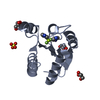
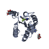
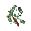
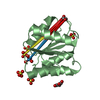
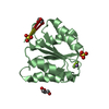
| Title | Structure of the CheYN59D/E89R Molybdate complex | ||||||
 Components Components | Chemotaxis protein CheY | ||||||
 Keywords Keywords | SIGNALING PROTEIN / two-component / signal transduction / response regulator / cheY / Beta-alpha protein / chemotaxis / CheZ / CheX / CheA / phosphorylation | ||||||
| Function / homology |  Function and homology information Function and homology informationbacterial-type flagellum basal body, C ring / bacterial-type flagellum rotor complex / bacterial-type flagellum-dependent swimming motility / aerotaxis / regulation of bacterial-type flagellum-dependent cell motility / regulation of chemotaxis / internal peptidyl-lysine acetylation / thermotaxis / bacterial-type flagellum / phosphorelay response regulator activity ...bacterial-type flagellum basal body, C ring / bacterial-type flagellum rotor complex / bacterial-type flagellum-dependent swimming motility / aerotaxis / regulation of bacterial-type flagellum-dependent cell motility / regulation of chemotaxis / internal peptidyl-lysine acetylation / thermotaxis / bacterial-type flagellum / phosphorelay response regulator activity / acetyltransferase activity / phosphorelay signal transduction system / chemotaxis / magnesium ion binding / signal transduction / cytoplasm / cytosol Similarity search - Function | ||||||
| Biological species |  | ||||||
| Method |  X-RAY DIFFRACTION / SYNCHROTRON / MOLECULAR REPLACEMENT / Resolution: 2.1 Å X-RAY DIFFRACTION / SYNCHROTRON / MOLECULAR REPLACEMENT / Resolution: 2.1 Å | ||||||
 Authors Authors | Immormino, R.M. / Starbird, C.A. / Silversmith, R.E. / Bourret, R.B. | ||||||
 Citation Citation | Journal: Biochemistry / Year: 2015 Title: Probing Mechanistic Similarities between Response Regulator Signaling Proteins and Haloacid Dehalogenase Phosphatases. Authors: Immormino, R.M. / Starbird, C.A. / Silversmith, R.E. / Bourret, R.B. | ||||||
| History |
|
- Structure visualization
| Structure viewer | Molecule: MolmilJmol/JSmol |
|---|
- Downloads & links
-Download
| PDBx/mmCIF format | 3rvr.cif.gz | 74.1 KB | Display | PDBx/mmCIF format |
|---|---|---|---|---|
| PDB format | pdb3rvr.ent.gz | 54.3 KB | Display | PDB format |
| PDBx/mmJSON format | 3rvr.json.gz | Tree view | PDBx/mmJSON format | |
| Others |  Other downloads Other downloads |
-Validation report
| Arichive directory | https://data.pdbj.org/pub/pdb/validation_reports/rv/3rvrftp://data.pdbj.org/pub/pdb/validation_reports/rv/3rvr | HTTPS FTP |
|---|
-Related structure data
| Related structure data | 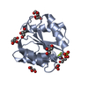 3rvjC 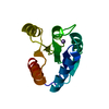 3rvkC 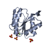 3rvlC  3rvmC 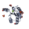 3rvnC  3rvoC 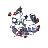 3rvpC 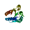 3rvqC 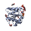 3rvsC 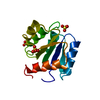 3chyS C: citing same article ( S: Starting model for refinement |
|---|---|
| Similar structure data |


-Links
 PDBj
PDBj

- Assembly
| Deposited unit | 
| ||||||||
|---|---|---|---|---|---|---|---|---|---|
| 1 | 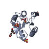
| ||||||||
| 2 | 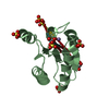
| ||||||||
| Unit cell |
|
-Components
-Protein , 1 types, 2 molecules AB
| #1: Protein | Mass: 14423.673 Da / Num. of mol.: 2 / Mutation: N59D, E89R Source method: isolated from a genetically manipulated source Source: (gene. exp.) |
|---|
-Non-polymers , 5 types, 308 molecules 


| #2: Chemical |  Mass: 54.938 Da / Num. of mol.: 2 / Source method: obtained synthetically / Formula: Mn Mass: 54.938 Da / Num. of mol.: 2 / Source method: obtained synthetically / Formula: Mn#3: Chemical |  Mass: 159.938 Da / Num. of mol.: 2 / Source method: obtained synthetically / Formula: MoO4 Mass: 159.938 Da / Num. of mol.: 2 / Source method: obtained synthetically / Formula: MoO4#4: Chemical | ChemComp-SO4 /  Mass: 96.063 Da / Num. of mol.: 11 / Source method: obtained synthetically / Formula: SO4 Mass: 96.063 Da / Num. of mol.: 11 / Source method: obtained synthetically / Formula: SO4#5: Chemical | ChemComp-GOL /  Mass: 92.094 Da / Num. of mol.: 5 / Source method: obtained synthetically / Formula: C3H8O3 Mass: 92.094 Da / Num. of mol.: 5 / Source method: obtained synthetically / Formula: C3H8O3#6: Water | ChemComp-HOH / | Mass: 18.015 Da / Num. of mol.: 288 / Source method: isolated from a natural source / Formula: H2O |
|---|
-Experimental details
-Experiment
| Experiment | Method: X-RAY DIFFRACTION / Number of used crystals: 1 |
|---|
- Sample preparation
| Crystal | Density Matthews: 4.02 Å3/Da / Density % sol: 69.44 % |
|---|---|
| Crystal grow | Temperature: 298 K / Method: vapor diffusion, hanging drop / pH: 7.5 Details: 2.45 M Ammonium Sulfate, 100 mM Tris, pH 7.5, 5% (v/v) Glycerol, 2 mM Ammonium Molybdate, 20mM Manganese chloride, 4.25 mg/mL CheY, VAPOR DIFFUSION, HANGING DROP, temperature 298K |
-Data collection
| Diffraction | Mean temperature: 100 K |
|---|---|
| Diffraction source | Source: SYNCHROTRON / Site: APS  / Beamline: 22-BM / Wavelength: 1.02631 Å / Beamline: 22-BM / Wavelength: 1.02631 Å |
| Detector | Type: MARMOSAIC 225 mm CCD / Detector: CCD / Date: Mar 27, 2011 |
| Radiation | Monochromator: sagital crystal / Protocol: SINGLE WAVELENGTH / Monochromatic (M) / Laue (L): M / Scattering type: x-ray |
| Radiation wavelength | Wavelength: 1.02631 Å / Relative weight: 1 |
| Reflection | Resolution: 2.1→50 Å / Num. all: 28023 / Num. obs: 27965 / % possible obs: 99.8 % / Observed criterion σ(I): -3 / Redundancy: 7 % / Biso Wilson estimate: 34.63 Å2 / Rsym value: 0.061 / Net I/σ(I): 20.2 |
| Reflection shell | Resolution: 2.1→2.15 Å / Redundancy: 6.7 % / Mean I/σ(I) obs: 5.33 / Num. unique all: 2038 / Rsym value: 0.282 / % possible all: 100 |
- Processing
| Software |
| ||||||||||||||||||||||||||||||||||||||||||||||||||||||||||||||||||||||||||||||||||||
|---|---|---|---|---|---|---|---|---|---|---|---|---|---|---|---|---|---|---|---|---|---|---|---|---|---|---|---|---|---|---|---|---|---|---|---|---|---|---|---|---|---|---|---|---|---|---|---|---|---|---|---|---|---|---|---|---|---|---|---|---|---|---|---|---|---|---|---|---|---|---|---|---|---|---|---|---|---|---|---|---|---|---|---|---|---|
| Refinement | Method to determine structure: MOLECULAR REPLACEMENT Starting model: PDB ENTRY 3CHY Resolution: 2.1→19.735 Å / SU ML: 0.22 / Isotropic thermal model: individual / Cross valid method: THROUGHOUT / σ(F): 0 / σ(I): -3 / Phase error: 16.55 / Stereochemistry target values: ML
| ||||||||||||||||||||||||||||||||||||||||||||||||||||||||||||||||||||||||||||||||||||
| Solvent computation | Shrinkage radii: 0.83 Å / VDW probe radii: 1.1 Å / Solvent model: FLAT BULK SOLVENT MODEL / Bsol: 44.312 Å2 / ksol: 0.347 e/Å3 | ||||||||||||||||||||||||||||||||||||||||||||||||||||||||||||||||||||||||||||||||||||
| Displacement parameters | Biso mean: 31.98 Å2
| ||||||||||||||||||||||||||||||||||||||||||||||||||||||||||||||||||||||||||||||||||||
| Refinement step | Cycle: LAST / Resolution: 2.1→19.735 Å
| ||||||||||||||||||||||||||||||||||||||||||||||||||||||||||||||||||||||||||||||||||||
| Refine LS restraints |
| ||||||||||||||||||||||||||||||||||||||||||||||||||||||||||||||||||||||||||||||||||||
| LS refinement shell |
|
