 Movie
Movie Controller

[English] 日本語
 Yorodumi
- PDB-1qir: ASPARTATE AMINOTRANSFERASE FROM ESCHERICHIA COLI, C191Y MUTATION,... -
+ Open data

- Basic information
| Entry | Database: PDB / ID: 1qir | ||||||
|---|---|---|---|---|---|---|---|
| Title | ASPARTATE AMINOTRANSFERASE FROM ESCHERICHIA COLI, C191Y MUTATION, WITH BOUND MALEATE | ||||||
 Components Components | ASPARTATE AMINOTRANSFERASE | ||||||
 Keywords Keywords | AMINOTRANSFERASE / TRANSFERASE(AMINOTRANSFERASE) / PYRIDOXAL PHOSPHATE / MALEATE | ||||||
| Function / homology |  Function and homology information Function and homology information: / L-tyrosine:2-oxoglutarate transaminase activity / L-phenylalanine biosynthetic process / aspartate transaminase / L-aspartate:2-oxoglutarate transaminase activity / pyridoxal phosphate binding / protein homodimerization activity / identical protein binding / cytosol / cytoplasm Similarity search - Function | ||||||
| Biological species |  | ||||||
| Method |  X-RAY DIFFRACTION / MOLECULAR REPLACEMENT / Resolution: 2.2 Å X-RAY DIFFRACTION / MOLECULAR REPLACEMENT / Resolution: 2.2 Å | ||||||
 Authors Authors | Jeffery, C.J. / Gloss, L.M. / Petsko, G.A. / Ringe, D. | ||||||
 Citation Citation | Journal: Protein Eng. / Year: 2000 Title: The Role of Residues Outside the Active Site in Catalysis: Structural Basis for Function of C191 Mutants of E. Coli Aspartate Aminotransferase Authors: Jeffery, C.J. / Gloss, L.M. / Petsko, G.A. / Ringe, D. | ||||||
| History |
|
- Structure visualization
| Structure viewer | Molecule: MolmilJmol/JSmol |
|---|
- Downloads & links
-Download
| PDBx/mmCIF format | 1qir.cif.gz | 106.6 KB | Display | PDBx/mmCIF format |
|---|---|---|---|---|
| PDB format | pdb1qir.ent.gz | 80.4 KB | Display | PDB format |
| PDBx/mmJSON format | 1qir.json.gz | Tree view | PDBx/mmJSON format | |
| Others |  Other downloads Other downloads |
-Validation report
| Arichive directory | https://data.pdbj.org/pub/pdb/validation_reports/qi/1qirftp://data.pdbj.org/pub/pdb/validation_reports/qi/1qir | HTTPS FTP |
|---|
-Related structure data
| Related structure data |  1b4xC  1qisC  1qitC 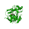 5eaaC  1asaS C: citing same article ( S: Starting model for refinement |
|---|---|
| Similar structure data |

-Links
 PDBj
PDBj- Assembly
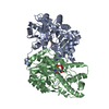
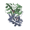
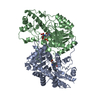
| Deposited unit | 
| ||||||||
|---|---|---|---|---|---|---|---|---|---|
| 1 | 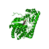
| ||||||||
| Unit cell |
| ||||||||
| Details | BIOLOGICAL_UNIT: ACTIVE AS A DIMER |
-Components
| #1: Protein | Mass: 43679.246 Da / Num. of mol.: 1 / Fragment: COMPLETE SUBUNIT / Mutation: YES Source method: isolated from a genetically manipulated source Details: PYRIDOXAL PHOSPHATE COFACTOR COVALENTLY BOUND TO LYS258 VIA SCHIFF BASE LINKAGE Source: (gene. exp.) |
|---|---|
| #2: Chemical | ChemComp-PLP /   Mass: 247.142 Da / Num. of mol.: 1 / Source method: obtained synthetically / Formula: C8H10NO6P Mass: 247.142 Da / Num. of mol.: 1 / Source method: obtained synthetically / Formula: C8H10NO6P |
| #3: Chemical | ChemComp-MAE /   Mass: 116.072 Da / Num. of mol.: 1 / Source method: obtained synthetically / Formula: C4H4O4 Mass: 116.072 Da / Num. of mol.: 1 / Source method: obtained synthetically / Formula: C4H4O4 |
| #4: Water | ChemComp-HOH /  Mass: 18.015 Da / Num. of mol.: 79 / Source method: isolated from a natural source / Formula: H2O Mass: 18.015 Da / Num. of mol.: 79 / Source method: isolated from a natural source / Formula: H2O |
| Compound details | NUMBERED TO MATCH NUMBERING OF CHICKEN CYTOPLASMI |
-Experimental details
-Experiment
| Experiment | Method: X-RAY DIFFRACTION / Number of used crystals: 1 |
|---|
- Sample preparation
| Crystal | Density Matthews: 3 Å3/Da / Density % sol: 59 % | ||||||||||||||||||||||||||||||||||||||||
|---|---|---|---|---|---|---|---|---|---|---|---|---|---|---|---|---|---|---|---|---|---|---|---|---|---|---|---|---|---|---|---|---|---|---|---|---|---|---|---|---|---|
| Crystal grow | pH: 7.5 Details: PROTEIN SOLUTION: 6MG/ML PROTEIN, 20 MM POTASSIUM PHOSPHATE BUFFER, PH 7.5, 10 UM PLP, 5 MM EDTA, RESERVOIR SOLUTION: 20MM POTASSIUM PHOSPHATE BUFFER, PH 7.5, AND 45-50% AMMONIUM SULFATE | ||||||||||||||||||||||||||||||||||||||||
| Crystal grow | *PLUS Method: vapor diffusion, hanging drop | ||||||||||||||||||||||||||||||||||||||||
| Components of the solutions | *PLUS
|
-Data collection
| Diffraction | Mean temperature: 297 K |
|---|---|
| Diffraction source | Source: ROTATING ANODE / Type: RIGAKU RUH2R / Wavelength: 1.5418 |
| Detector | Type: RIGAKU IMAGE PLATE / Detector: IMAGE PLATE |
| Radiation | Monochromator: NI FILTER / Protocol: SINGLE WAVELENGTH / Monochromatic (M) / Laue (L): M / Scattering type: x-ray |
| Radiation wavelength | Wavelength: 1.5418 Å / Relative weight: 1 |
| Reflection | Resolution: 2.2→50 Å / Num. obs: 34520 / % possible obs: 80 % / Observed criterion σ(I): 0 / Redundancy: 1 % / Rmerge(I) obs: 0.06 |
| Reflection shell | Highest resolution: 2.2 Å |
- Processing
| Software |
| ||||||||||||||||||||||||||||||||||||||||||||||||||||||||||||
|---|---|---|---|---|---|---|---|---|---|---|---|---|---|---|---|---|---|---|---|---|---|---|---|---|---|---|---|---|---|---|---|---|---|---|---|---|---|---|---|---|---|---|---|---|---|---|---|---|---|---|---|---|---|---|---|---|---|---|---|---|---|
| Refinement | Method to determine structure: MOLECULAR REPLACEMENT Starting model: 1ASA Resolution: 2.2→10 Å / Data cutoff high absF: 1000000 / Data cutoff low absF: 0 / Cross valid method: THROUGHOUT / σ(F): 0
| ||||||||||||||||||||||||||||||||||||||||||||||||||||||||||||
| Displacement parameters | Biso mean: 25.09 Å2 | ||||||||||||||||||||||||||||||||||||||||||||||||||||||||||||
| Refinement step | Cycle: LAST / Resolution: 2.2→10 Å
| ||||||||||||||||||||||||||||||||||||||||||||||||||||||||||||
| Refine LS restraints |
| ||||||||||||||||||||||||||||||||||||||||||||||||||||||||||||
| Xplor file | Serial no: 1 / Param file: PARHCSDX.PRO / Topol file: TOPHCSDX.PRO | ||||||||||||||||||||||||||||||||||||||||||||||||||||||||||||
| Software | *PLUS Name: X-PLOR / Version: 3.851 / Classification: refinement | ||||||||||||||||||||||||||||||||||||||||||||||||||||||||||||
| Refine LS restraints | *PLUS
|
