 Movie
Movie Controller


+ Open data

- Basic information
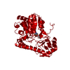
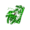
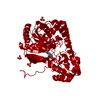
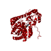
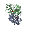
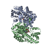
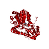
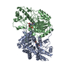
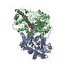
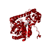
| Entry | Database: PDB / ID: 1ahg | |||||||||
|---|---|---|---|---|---|---|---|---|---|---|
| Title | ASPARTATE AMINOTRANSFERASE HEXAMUTANT | |||||||||
 Components Components | Aspartate aminotransferase | |||||||||
 Keywords Keywords | TRANSFERASE (AMINOTRANSFERASE) | |||||||||
| Function / homology |  Function and homology information Function and homology information: / L-tyrosine:2-oxoglutarate transaminase activity / L-phenylalanine biosynthetic process / aspartate transaminase / L-aspartate:2-oxoglutarate transaminase activity / pyridoxal phosphate binding / protein homodimerization activity / identical protein binding / cytosol / cytoplasm Similarity search - Function | |||||||||
| Biological species |  | |||||||||
| Method |  X-RAY DIFFRACTION / Resolution: 2.5 Å X-RAY DIFFRACTION / Resolution: 2.5 Å | |||||||||
 Authors Authors | Malashkevich, V.N. / Jansonius, J.N. | |||||||||
 Citation Citation | Journal: Nat.Struct.Biol. / Year: 1995 Title: Alternating arginine-modulated substrate specificity in an engineered tyrosine aminotransferase. Authors: Malashkevich, V.N. / Onuffer, J.J. / Kirsch, J.F. / Jansonius, J.N. #1: Journal: Biochemistry of Vitamin B6 and Pqq / Year: 1994Title: Redesign of Aspartate Aminotransferase Specificity to that of Tyrosine Aminotransferase Authors: Kirsch, J.F. / Onuffer, J.J. | |||||||||
| History |
|
- Structure visualization
| Structure viewer | Molecule: MolmilJmol/JSmol |
|---|
- Downloads & links
-Download
| PDBx/mmCIF format | 1ahg.cif.gz | 177.7 KB | Display | PDBx/mmCIF format |
|---|---|---|---|---|
| PDB format | pdb1ahg.ent.gz | 139.8 KB | Display | PDB format |
| PDBx/mmJSON format | 1ahg.json.gz | Tree view | PDBx/mmJSON format | |
| Others |  Other downloads Other downloads |
-Validation report
| Arichive directory | https://data.pdbj.org/pub/pdb/validation_reports/ah/1ahgftp://data.pdbj.org/pub/pdb/validation_reports/ah/1ahg | HTTPS FTP |
|---|
-Related structure data


-Links
 PDBj
PDBj

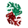
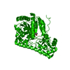
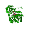
- Assembly
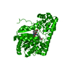
| Deposited unit | 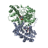
| ||||||||
|---|---|---|---|---|---|---|---|---|---|
| 1 |
| ||||||||
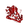
| Unit cell |
| ||||||||
| Atom site foot note | 1: CIS PROLINE - PRO A 138 / 2: CIS PROLINE - PRO A 195 / 3: CIS PROLINE - PRO B 138 / 4: CIS PROLINE - PRO B 195 | ||||||||
| Noncrystallographic symmetry (NCS) | NCS oper: (Code: given Matrix: (-1, 0.00039, 0.00101), Vector: Details | MTRIX THE TRANSFORMATIONS PRESENTED ON MTRIX RECORDS BELOW DESCRIBE NON-CRYSTALLOGRAPHIC RELATIONSHIPS AMONG THE VARIOUS DOMAINS IN THIS ENTRY. APPLYING THE APPROPRIATE MTRIX TRANSFORMATION TO THE RESIDUES LISTED FIRST WILL YIELD APPROXIMATE COORDINATES FOR THE RESIDUES LISTED SECOND. APPLIED TO TRANSFORMED TO MTRIX RESIDUES RESIDUES RMSD M1 A 5 .. A 409 B 5 .. B 409 0.265 | |
-Components
| #1: Protein | Mass: 43637.293 Da / Num. of mol.: 2 Source method: isolated from a genetically manipulated source Source: (gene. exp.) Strain: K12 / Gene: aspC, b0928, JW0911 / Production host: #2: Chemical |   Type: L-peptide linking / Mass: 181.189 Da / Num. of mol.: 2 / Source method: obtained synthetically / Formula: C9H11NO3 Type: L-peptide linking / Mass: 181.189 Da / Num. of mol.: 2 / Source method: obtained synthetically / Formula: C9H11NO3#3: Chemical |   Mass: 247.142 Da / Num. of mol.: 2 / Source method: obtained synthetically / Formula: C8H10NO6P Mass: 247.142 Da / Num. of mol.: 2 / Source method: obtained synthetically / Formula: C8H10NO6P#4: Water | ChemComp-HOH / |  Mass: 18.015 Da / Num. of mol.: 510 / Source method: isolated from a natural source / Formula: H2O Mass: 18.015 Da / Num. of mol.: 510 / Source method: isolated from a natural source / Formula: H2OCompound details | COMPND SUBSTRATE SPECIFICIT | |
|---|
-Experimental details
-Experiment
| Experiment | Method: X-RAY DIFFRACTION |
|---|
- Sample preparation
| Crystal | Density Matthews: 3.03 Å3/Da / Density % sol: 59.46 % | |||||||||||||||||||||||||||||||||||||||||||||
|---|---|---|---|---|---|---|---|---|---|---|---|---|---|---|---|---|---|---|---|---|---|---|---|---|---|---|---|---|---|---|---|---|---|---|---|---|---|---|---|---|---|---|---|---|---|---|
| Crystal grow | *PLUS pH: 7.5 / Method: vapor diffusion, hanging drop | |||||||||||||||||||||||||||||||||||||||||||||
| Components of the solutions | *PLUS
|
-Data collection
| Diffraction source | Wavelength: 1.5418 Å |
|---|---|
| Detector | Type: MARRESEARCH / Detector: IMAGE PLATE / Date: Apr 4, 1993 |
| Radiation | Monochromatic (M) / Laue (L): M / Scattering type: x-ray |
| Radiation wavelength | Wavelength: 1.5418 Å / Relative weight: 1 |
| Reflection | Redundancy: 3.2 % / Biso Wilson estimate: 33.5 Å2 |
| Reflection | *PLUS Num. obs: 33157 / % possible obs: 90.5 % / Observed criterion σ(I): 1 |
- Processing
| Software |
| ||||||||||||||||||||||||||||||||||||||||||||||||||
|---|---|---|---|---|---|---|---|---|---|---|---|---|---|---|---|---|---|---|---|---|---|---|---|---|---|---|---|---|---|---|---|---|---|---|---|---|---|---|---|---|---|---|---|---|---|---|---|---|---|---|---|
| Refinement | Resolution: 2.5→8 Å / σ(F): 1 /
| ||||||||||||||||||||||||||||||||||||||||||||||||||
| Refinement step | Cycle: LAST / Resolution: 2.5→8 Å
| ||||||||||||||||||||||||||||||||||||||||||||||||||
| Refine LS restraints |
|
