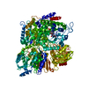
 Movie
Movie Controller


+ Open data

- Basic information
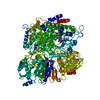
| Entry | Database: PDB / ID: 1qfx | |||||||||
|---|---|---|---|---|---|---|---|---|---|---|
| Title | PH 2.5 ACID PHOSPHATASE FROM ASPERGILLUS NIGER | |||||||||
 Components Components | PROTEIN (PH 2.5 ACID PHOSPHATASE) | |||||||||
 Keywords Keywords | HYDROLASE / PHOSPHOMONOESTERASE | |||||||||
| Function / homology |  Function and homology information Function and homology information3-phytase / inositol hexakisphosphate 3-phosphatase activity / acid phosphatase activity / fungal-type cell wall Similarity search - Function | |||||||||
| Biological species |  | |||||||||
| Method |  X-RAY DIFFRACTION / SYNCHROTRON / SIRAS / Resolution: 2.4 Å X-RAY DIFFRACTION / SYNCHROTRON / SIRAS / Resolution: 2.4 Å | |||||||||
 Authors Authors | Kostrewa, D. / Wyss, M. / D'Arcy, A. / Van Loon, A.P.G.M. | |||||||||
 Citation Citation | Journal: J.Mol.Biol. / Year: 1999 Title: Crystal structure of Aspergillus niger pH 2.5 acid phosphatase at 2. 4 A resolution. Authors: Kostrewa, D. / Wyss, M. / D'Arcy, A. / van Loon, A.P. | |||||||||
| History |
|
- Structure visualization
| Structure viewer | Molecule: MolmilJmol/JSmol |
|---|
- Downloads & links
-Download
| PDBx/mmCIF format | 1qfx.cif.gz | 203.3 KB | Display | PDBx/mmCIF format |
|---|---|---|---|---|
| PDB format | pdb1qfx.ent.gz | 161.3 KB | Display | PDB format |
| PDBx/mmJSON format | 1qfx.json.gz | Tree view | PDBx/mmJSON format | |
| Others |  Other downloads Other downloads |
-Validation report
| Arichive directory | https://data.pdbj.org/pub/pdb/validation_reports/qf/1qfxftp://data.pdbj.org/pub/pdb/validation_reports/qf/1qfx | HTTPS FTP |
|---|
-Related structure data
| Similar structure data |
|---|


-Links
 PDBj
PDBj


- Assembly
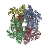
| Deposited unit | 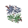
| |||||||||||
|---|---|---|---|---|---|---|---|---|---|---|---|---|
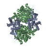
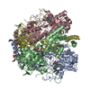
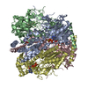
| 1 | 
| |||||||||||
| Unit cell |
| |||||||||||
| Noncrystallographic symmetry (NCS) | NCS domain:
NCS oper: (Code: given Matrix: (-0.97673, 0.03956, 0.21077), Vector: Details | THE ASYMMETRIC UNIT OF THE CRYSTAL CONTAINS A DIMER, RELATED BY A LOCAL DYAD. THIS DIMER FORMS A TETRAMER WITH ALMOST PROPER 222 (D2) SYMMETRY BY A CRYSTALLOGRAPHIC DYAD. | |
-Components
-Protein , 1 types, 2 molecules AB
| #1: Protein | Mass: 50879.887 Da / Num. of mol.: 2 / Source method: isolated from a natural source Details: N-GLYCOSYLATED AT A 172, A 296, A439, B 172, B 296 AND B 439 Source: (natural) |
|---|
-Sugars , 3 types, 6 molecules 
| #2: Polysaccharide | alpha-D-mannopyranose-(1-2)-alpha-D-mannopyranose-(1-2)-alpha-D-mannopyranose-(1-3)-[alpha-D- ...alpha-D-mannopyranose-(1-2)-alpha-D-mannopyranose-(1-2)-alpha-D-mannopyranose-(1-3)-[alpha-D-mannopyranose-(1-6)]beta-D-mannopyranose-(1-4)-2-acetamido-2-deoxy-beta-D-glucopyranose-(1-4)-2-acetamido-2-deoxy-beta-D-glucopyranose Source method: isolated from a genetically manipulated source |
|---|---|
| #3: Polysaccharide | alpha-D-mannopyranose-(1-3)-[alpha-D-mannopyranose-(1-6)]beta-D-mannopyranose-(1-4)-2-acetamido-2- ...alpha-D-mannopyranose-(1-3)-[alpha-D-mannopyranose-(1-6)]beta-D-mannopyranose-(1-4)-2-acetamido-2-deoxy-beta-D-glucopyranose-(1-4)-2-acetamido-2-deoxy-beta-D-glucopyranose Source method: isolated from a genetically manipulated source |
| #4: Sugar | ChemComp-NAG /  Type: D-saccharide, beta linking / Mass: 221.208 Da / Num. of mol.: 4 Type: D-saccharide, beta linking / Mass: 221.208 Da / Num. of mol.: 4Source method: isolated from a genetically manipulated source Formula: C8H15NO6 |
-Non-polymers , 3 types, 665 molecules 


| #5: Chemical | ChemComp-SO4 /  Mass: 96.063 Da / Num. of mol.: 11 / Source method: obtained synthetically / Formula: SO4 Mass: 96.063 Da / Num. of mol.: 11 / Source method: obtained synthetically / Formula: SO4#6: Chemical | ChemComp-GOL /  Mass: 92.094 Da / Num. of mol.: 6 / Source method: obtained synthetically / Formula: C3H8O3 Mass: 92.094 Da / Num. of mol.: 6 / Source method: obtained synthetically / Formula: C3H8O3#7: Water | ChemComp-HOH / | Mass: 18.015 Da / Num. of mol.: 648 / Source method: isolated from a natural source / Formula: H2O |
|---|
-Details
| Has protein modification | Y |
|---|
-Experimental details
-Experiment
| Experiment | Method: X-RAY DIFFRACTION / Number of used crystals: 1 |
|---|
- Sample preparation
| Crystal | Density Matthews: 4.2 Å3/Da / Density % sol: 70.4 % | ||||||||||||||||||||
|---|---|---|---|---|---|---|---|---|---|---|---|---|---|---|---|---|---|---|---|---|---|
| Crystal grow | pH: 7 / Details: 0.1M HEPES/HCL PH 7.0, 2.4M AMMONIUM SULFATE | ||||||||||||||||||||
| Crystal | *PLUS | ||||||||||||||||||||
| Crystal grow | *PLUS Method: vapor diffusion, hanging drop | ||||||||||||||||||||
| Components of the solutions | *PLUS
|
-Data collection
| Diffraction | Mean temperature: 120 K |
|---|---|
| Diffraction source | Source: SYNCHROTRON / Site: ESRF  / Beamline: BM1A / Wavelength: 0.873 / Beamline: BM1A / Wavelength: 0.873 |
| Detector | Type: MARRESEARCH / Detector: IMAGE PLATE / Date: Jan 29, 1997 / Details: MIRROR |
| Radiation | Monochromator: DOUBLE CRYSTAL / Protocol: SINGLE WAVELENGTH / Monochromatic (M) / Laue (L): M / Scattering type: x-ray |
| Radiation wavelength | Wavelength: 0.873 Å / Relative weight: 1 |
| Reflection | Resolution: 2.4→25 Å / Num. obs: 58656 / % possible obs: 86.5 % / Observed criterion σ(I): 0 / Redundancy: 2.9 % / Biso Wilson estimate: 37.3 Å2 / Rsym value: 0.056 / Net I/σ(I): 18.8 |
| Reflection shell | Resolution: 2.4→2.5 Å / Redundancy: 1.7 % / Mean I/σ(I) obs: 4.2 / Rsym value: 0.182 / % possible all: 67.3 |
| Reflection | *PLUS Num. measured all: 173451 / Rmerge(I) obs: 0.055 |
| Reflection shell | *PLUS % possible obs: 67.3 % / Rmerge(I) obs: 0.182 |
- Processing
| Software |
| ||||||||||||||||||||||||||||||||||||||||||||||||||||||||||||
|---|---|---|---|---|---|---|---|---|---|---|---|---|---|---|---|---|---|---|---|---|---|---|---|---|---|---|---|---|---|---|---|---|---|---|---|---|---|---|---|---|---|---|---|---|---|---|---|---|---|---|---|---|---|---|---|---|---|---|---|---|---|
| Refinement | Method to determine structure: SIRAS / Resolution: 2.4→25 Å / Data cutoff high absF: 100000 / Data cutoff low absF: 0 / Isotropic thermal model: RESTRAINED / Cross valid method: FREE-R / σ(F): 0 Details: BULK SOLVENT CORRECTION WITH PROBE=1.0A,SHRINK=1.1A,K3=0.37E/A**3,B3=35.7A**2.
| ||||||||||||||||||||||||||||||||||||||||||||||||||||||||||||
| Displacement parameters | Biso mean: 25.6 Å2 | ||||||||||||||||||||||||||||||||||||||||||||||||||||||||||||
| Refine analyze | Luzzati d res low obs: 25 Å / Luzzati sigma a obs: 0.3 Å | ||||||||||||||||||||||||||||||||||||||||||||||||||||||||||||
| Refinement step | Cycle: LAST / Resolution: 2.4→25 Å
| ||||||||||||||||||||||||||||||||||||||||||||||||||||||||||||
| Refine LS restraints |
| ||||||||||||||||||||||||||||||||||||||||||||||||||||||||||||
| Refine LS restraints NCS |
| ||||||||||||||||||||||||||||||||||||||||||||||||||||||||||||
| LS refinement shell | Resolution: 2.4→2.42 Å / Total num. of bins used: 50
| ||||||||||||||||||||||||||||||||||||||||||||||||||||||||||||
| Xplor file |
| ||||||||||||||||||||||||||||||||||||||||||||||||||||||||||||
| Software | *PLUS Name: X-PLOR / Version: 3.851 / Classification: refinement | ||||||||||||||||||||||||||||||||||||||||||||||||||||||||||||
| Refinement | *PLUS Highest resolution: 2.4 Å / Lowest resolution: 25 Å / σ(F): 0 / % reflection Rfree: 5 % / Rfactor obs: 0.18 | ||||||||||||||||||||||||||||||||||||||||||||||||||||||||||||
| Solvent computation | *PLUS | ||||||||||||||||||||||||||||||||||||||||||||||||||||||||||||
| Displacement parameters | *PLUS Biso mean: 25.6 Å2 | ||||||||||||||||||||||||||||||||||||||||||||||||||||||||||||
| Refine LS restraints | *PLUS
| ||||||||||||||||||||||||||||||||||||||||||||||||||||||||||||
| LS refinement shell | *PLUS Rfactor Rfree: 0.329 / % reflection Rfree: 5 % / Rfactor Rwork: 0.302 |
