 Movie
Movie Controller


+ Open data

- Basic information
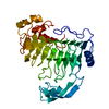
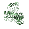
| Entry | Database: PDB / ID: 1qcx | ||||||
|---|---|---|---|---|---|---|---|
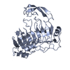
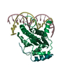
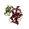
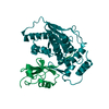
| Title | PECTIN LYASE B | ||||||
 Components Components | PECTIN LYASE B | ||||||
 Keywords Keywords | LYASE / PECTIN LYASE / BETA-HELIX PROTEIN / PECTIN / PLANT CELL WALL | ||||||
| Function / homology |  Function and homology information Function and homology informationpectin lyase / pectin lyase activity / pectate lyase activity / polysaccharide catabolic process / cell wall organization / extracellular region Similarity search - Function | ||||||
| Biological species |  | ||||||
| Method |  X-RAY DIFFRACTION / MOLECULAR REPLACEMENT, HEAVY ATOMS / Resolution: 1.7 Å X-RAY DIFFRACTION / MOLECULAR REPLACEMENT, HEAVY ATOMS / Resolution: 1.7 Å | ||||||
 Authors Authors | Vitali, J. / Jurnak, F. | ||||||
 Citation Citation | Journal: Plant Physiol. / Year: 1998 Title: The tree-dimensional structure of aspergillus niger pectin lyase B at 1.7-A resolution. Authors: Vitali, J. / Schick, B. / Kester, H.C. / Visser, J. / Jurnak, F. | ||||||
| History |
|
- Structure visualization
| Structure viewer | Molecule: MolmilJmol/JSmol |
|---|
- Downloads & links
-Download
| PDBx/mmCIF format | 1qcx.cif.gz | 84.7 KB | Display | PDBx/mmCIF format |
|---|---|---|---|---|
| PDB format | pdb1qcx.ent.gz | 63.1 KB | Display | PDB format |
| PDBx/mmJSON format | 1qcx.json.gz | Tree view | PDBx/mmJSON format | |
| Others |  Other downloads Other downloads |
-Validation report
| Arichive directory | https://data.pdbj.org/pub/pdb/validation_reports/qc/1qcxftp://data.pdbj.org/pub/pdb/validation_reports/qc/1qcx | HTTPS FTP |
|---|
-Related structure data
| Similar structure data |
|---|

-Links
 PDBj
PDBj- Assembly
| Deposited unit | 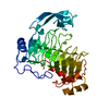
| ||||||||
|---|---|---|---|---|---|---|---|---|---|
| 1 |
| ||||||||
| Unit cell |
|
-Components
| #1: Protein | Mass: 37837.914 Da / Num. of mol.: 1 / Source method: isolated from a natural source / Source: (natural) |
|---|---|
| #2: Water | ChemComp-HOH /  Mass: 18.015 Da / Num. of mol.: 339 / Source method: isolated from a natural source / Formula: H2O Mass: 18.015 Da / Num. of mol.: 339 / Source method: isolated from a natural source / Formula: H2O |
| Has protein modification | Y |
-Experimental details
-Experiment
| Experiment | Method: X-RAY DIFFRACTION / Number of used crystals: 1 |
|---|
- Sample preparation
| Crystal | Density Matthews: 2.07 Å3/Da / Density % sol: 41 % | |||||||||||||||||||||||||
|---|---|---|---|---|---|---|---|---|---|---|---|---|---|---|---|---|---|---|---|---|---|---|---|---|---|---|
| Crystal grow | pH: 5 / Details: pH 5.0 | |||||||||||||||||||||||||
| Crystal | *PLUS Density % sol: 41 % | |||||||||||||||||||||||||
| Crystal grow | *PLUS Temperature: 4 ℃ / pH: 7.5 / Method: vapor diffusion | |||||||||||||||||||||||||
| Components of the solutions | *PLUS
|
-Data collection
| Diffraction | Mean temperature: 295 K |
|---|---|
| Diffraction source | Source: ROTATING ANODE / Type: RIGAKU RUH2R / Wavelength: 1.5418 |
| Detector | Type: XUONG-HAMLIN MULTIWIRE / Detector: AREA DETECTOR |
| Radiation | Monochromator: GRAPHITE(002) / Protocol: SINGLE WAVELENGTH / Monochromatic (M) / Laue (L): M / Scattering type: x-ray |
| Radiation wavelength | Wavelength: 1.5418 Å / Relative weight: 1 |
| Reflection | Resolution: 1.7→100 Å / Num. obs: 50124 / % possible obs: 73.8 % / Observed criterion σ(I): 0 / Redundancy: 4.1 % / Rsym value: 0.054 / Net I/σ(I): 18.9 |
| Reflection | *PLUS Num. measured all: 204035 / Rmerge(I) obs: 0.054 |
- Processing
| Software |
| ||||||||||||||||||||||||||||||||||||||||||||||||||||||||||||
|---|---|---|---|---|---|---|---|---|---|---|---|---|---|---|---|---|---|---|---|---|---|---|---|---|---|---|---|---|---|---|---|---|---|---|---|---|---|---|---|---|---|---|---|---|---|---|---|---|---|---|---|---|---|---|---|---|---|---|---|---|---|
| Refinement | Method to determine structure: MOLECULAR REPLACEMENT, HEAVY ATOMS Starting model: FOR MOLECULAR REPLACEMENT: PARALLEL BETA HELIX OF PELE, ALL AMINO ACIDS REPLACED WITH ALA UNLESS COMMON IN THE TWO PROTEINS; HEAVY ATOMS: PT, HG Resolution: 1.7→7 Å / Rfactor Rfree error: 0.003 / Data cutoff high absF: 100000 / Data cutoff low absF: 0.1 / Cross valid method: R-FREE / σ(F): 2 Details: FREE R WAS USED UNTIL LAST CYCLE WHEN ALL DATA WAS INCLUDED
| ||||||||||||||||||||||||||||||||||||||||||||||||||||||||||||
| Displacement parameters | Biso mean: 17.5 Å2 | ||||||||||||||||||||||||||||||||||||||||||||||||||||||||||||
| Refinement step | Cycle: LAST / Resolution: 1.7→7 Å
| ||||||||||||||||||||||||||||||||||||||||||||||||||||||||||||
| Refine LS restraints |
| ||||||||||||||||||||||||||||||||||||||||||||||||||||||||||||
| LS refinement shell | Resolution: 1.78→1.87 Å / Rfactor Rfree error: 0.01 / Total num. of bins used: 8
| ||||||||||||||||||||||||||||||||||||||||||||||||||||||||||||
| Software | *PLUS Name: X-PLOR / Version: 3.1 / Classification: refinement | ||||||||||||||||||||||||||||||||||||||||||||||||||||||||||||
| Refine LS restraints | *PLUS
|
