 Movie
Movie Controller


+ Open data

- Basic information
| Entry | Database: PDB / ID: 1idk | ||||||
|---|---|---|---|---|---|---|---|
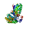
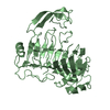
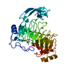
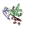
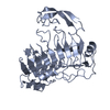
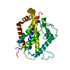
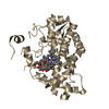
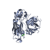
| Title | PECTIN LYASE A | ||||||
 Components Components | PECTIN LYASE A | ||||||
 Keywords Keywords | LYASE / GLYCOPROTEIN / MULTIGENE FAMILY | ||||||
| Function / homology |  Function and homology information Function and homology informationpectin lyase / pectin lyase activity / pectate lyase activity / polysaccharide catabolic process / cell wall organization / extracellular region Similarity search - Function | ||||||
| Biological species |  | ||||||
| Method |  X-RAY DIFFRACTION / SYNCHROTRON / molecular replacement, MIR / Resolution: 1.93 Å X-RAY DIFFRACTION / SYNCHROTRON / molecular replacement, MIR / Resolution: 1.93 Å | ||||||
 Authors Authors | Mayans, O. / Scott, M. / Connerton, I. / Gravesen, T. / Benen, J. / Visser, J. / Pickersgill, R. / Jenkins, J. | ||||||
 Citation Citation | Journal: Structure / Year: 1997 Title: Two crystal structures of pectin lyase A from Aspergillus reveal a pH driven conformational change and striking divergence in the substrate-binding clefts of pectin and pectate lyases. Authors: Mayans, O. / Scott, M. / Connerton, I. / Gravesen, T. / Benen, J. / Visser, J. / Pickersgill, R. / Jenkins, J. #1: Journal: Acta Crystallogr.,Sect.D / Year: 1996Title: Crystallization and Preliminary X-Ray Analysis of Pectin Lyase a from Aspergillus Niger Authors: Jenkins, J. / Scott, M. / Mayans, O. / Pickersgill, R. / Harris, G. / Connerton, I. / Gravesen, T. #2: Journal: Nat.Struct.Biol. / Year: 1994Title: The Structure of Bacillus Subtilis Pectate Lyase in Complex with Calcium Authors: Pickersgill, R. / Jenkins, J. / Harris, G. / Nasser, W. / Robert-Baudouy, J. | ||||||
| History |
|
- Structure visualization
| Structure viewer | Molecule: MolmilJmol/JSmol |
|---|
- Downloads & links
-Download
| PDBx/mmCIF format | 1idk.cif.gz | 83.3 KB | Display | PDBx/mmCIF format |
|---|---|---|---|---|
| PDB format | pdb1idk.ent.gz | 61.9 KB | Display | PDB format |
| PDBx/mmJSON format | 1idk.json.gz | Tree view | PDBx/mmJSON format | |
| Others |  Other downloads Other downloads |
-Validation report
| Arichive directory | https://data.pdbj.org/pub/pdb/validation_reports/id/1idkftp://data.pdbj.org/pub/pdb/validation_reports/id/1idk | HTTPS FTP |
|---|
-Related structure data


-Links
 PDBj
PDBj- Assembly
| Deposited unit | 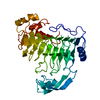
| ||||||||
|---|---|---|---|---|---|---|---|---|---|
| 1 |
| ||||||||
| Unit cell |
|
-Components
| #1: Protein | Mass: 37982.188 Da / Num. of mol.: 1 / Source method: isolated from a natural source / Details: SECRETED PROTEIN / Source: (natural) |
|---|---|
| #2: Water | ChemComp-HOH /  Mass: 18.015 Da / Num. of mol.: 175 / Source method: isolated from a natural source / Formula: H2O Mass: 18.015 Da / Num. of mol.: 175 / Source method: isolated from a natural source / Formula: H2O |
| Has protein modification | Y |
-Experimental details
-Experiment
| Experiment | Method: X-RAY DIFFRACTION / Number of used crystals: 1 |
|---|
- Sample preparation
| Crystal | Density Matthews: 2.3 Å3/Da / Density % sol: 47 % | ||||||||||||||||||||||||||||||
|---|---|---|---|---|---|---|---|---|---|---|---|---|---|---|---|---|---|---|---|---|---|---|---|---|---|---|---|---|---|---|---|
| Crystal grow | pH: 8.5 Details: 28% PEG 4000, 100 MM SODIUM ACETATE, 100 MM TRIS-HCL AT PH 8.5 | ||||||||||||||||||||||||||||||
| Crystal grow | *PLUS Method: vapor diffusion, hanging dropDetails: Jenkins, J., (1996) Acta Crystallogr.,Sect.D, 52, 402. | ||||||||||||||||||||||||||||||
| Components of the solutions | *PLUS
|
-Data collection
| Diffraction | Mean temperature: 290 K |
|---|---|
| Diffraction source | Source: SYNCHROTRON / Site: SRS  / Beamline: PX9.6 / Wavelength: 0.87 / Beamline: PX9.6 / Wavelength: 0.87 |
| Detector | Type: MARRESEARCH / Detector: IMAGE PLATE / Date: Jun 9, 1994 |
| Radiation | Monochromatic (M) / Laue (L): M / Scattering type: x-ray |
| Radiation wavelength | Wavelength: 0.87 Å / Relative weight: 1 |
| Reflection | Resolution: 1.93→25.44 Å / Num. obs: 25695 / % possible obs: 94 % / Redundancy: 3.1 % / Biso Wilson estimate: 23.66 Å2 / Rmerge(I) obs: 0.061 / Net I/σ(I): 19.47 |
| Reflection shell | Resolution: 1.93→1.97 Å / Redundancy: 2.2 % / Rmerge(I) obs: 0.25 / Mean I/σ(I) obs: 5.3 / % possible all: 72.3 |
| Reflection | *PLUS Num. measured all: 80561 |
| Reflection shell | *PLUS % possible obs: 84.3 % |
- Processing
| Software |
| ||||||||||||||||||||||||||||||||||||||||||||||||||||||||||||||||||||||||||||||||
|---|---|---|---|---|---|---|---|---|---|---|---|---|---|---|---|---|---|---|---|---|---|---|---|---|---|---|---|---|---|---|---|---|---|---|---|---|---|---|---|---|---|---|---|---|---|---|---|---|---|---|---|---|---|---|---|---|---|---|---|---|---|---|---|---|---|---|---|---|---|---|---|---|---|---|---|---|---|---|---|---|---|
| Refinement | Method to determine structure: molecular replacement, MIR Starting model: DERIVED FROM BACILLUS SUBTILIS PECTATE LYASE Resolution: 1.93→15 Å / Cross valid method: FREE R + R VALUE OF SECOND CRYSTAL FORM / σ(F): 2 Details: OVERALL ANISOTROPIC SCALING, BULK SOLVENT CORRECTION AND RESTRAINED ISOTROPIC INDIVIDUAL TEMPERATURE FACTOR REFINEMENT
| ||||||||||||||||||||||||||||||||||||||||||||||||||||||||||||||||||||||||||||||||
| Displacement parameters | Biso mean: 32.7 Å2
| ||||||||||||||||||||||||||||||||||||||||||||||||||||||||||||||||||||||||||||||||
| Refinement step | Cycle: LAST / Resolution: 1.93→15 Å
| ||||||||||||||||||||||||||||||||||||||||||||||||||||||||||||||||||||||||||||||||
| Refine LS restraints |
| ||||||||||||||||||||||||||||||||||||||||||||||||||||||||||||||||||||||||||||||||
| LS refinement shell | Resolution: 1.93→1.97 Å
| ||||||||||||||||||||||||||||||||||||||||||||||||||||||||||||||||||||||||||||||||
| Xplor file |
| ||||||||||||||||||||||||||||||||||||||||||||||||||||||||||||||||||||||||||||||||
| Software | *PLUS Name: X-PLOR / Version: 3.1 / Classification: refinement | ||||||||||||||||||||||||||||||||||||||||||||||||||||||||||||||||||||||||||||||||
| Refine LS restraints | *PLUS
|
