 Movie
Movie Controller


+ Open data

- Basic information
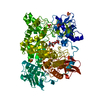
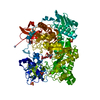
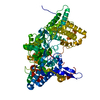
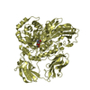
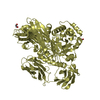
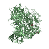
| Entry | Database: PDB / ID: 1qba | ||||||
|---|---|---|---|---|---|---|---|
| Title | BACTERIAL CHITOBIASE, GLYCOSYL HYDROLASE FAMILY 20 | ||||||
 Components Components | CHITOBIASE | ||||||
 Keywords Keywords | GLYCOSYL HYDROLASE / CHITOBIASE / CHITINOLYSIS / BA8-BARREL | ||||||
| Function / homology |  Function and homology information Function and homology informationglycosaminoglycan metabolic process / beta-N-acetylhexosaminidase activity / beta-N-acetylhexosaminidase / chitin catabolic process / polysaccharide binding / polysaccharide catabolic process / periplasmic space / membrane Similarity search - Function | ||||||
| Biological species |  Serratia marcescens (bacteria) Serratia marcescens (bacteria) | ||||||
| Method |  X-RAY DIFFRACTION / SYNCHROTRON / MIR / Resolution: 1.85 Å X-RAY DIFFRACTION / SYNCHROTRON / MIR / Resolution: 1.85 Å | ||||||
 Authors Authors | Tews, I. / Perrakis, A. / Oppenheim, A. / Dauter, Z. / Wilson, K.S. / Vorgias, C.E. | ||||||
 Citation Citation | Journal: Nat.Struct.Biol. / Year: 1996 Title: Bacterial chitobiase structure provides insight into catalytic mechanism and the basis of Tay-Sachs disease. Authors: Tews, I. / Perrakis, A. / Oppenheim, A. / Dauter, Z. / Wilson, K.S. / Vorgias, C.E. #1: Journal: Gene / Year: 1996Title: N-Acetylglucosaminidase (Chitobiase) from Serratia Marcescens: Gene Sequence, and Protein Production and Purification in Escherichia Coli Authors: Tews, I. / Vincentelli, R. / Vorgias, C.E. #3: Journal: Mol.Gen.Genet. / Year: 1989Title: Cloning of the Gene Coding for Chitobiase of Serratia Marcescens Authors: Kless, H. / Sitrit, Y. / Chet, I. / Oppenheim, A.B. | ||||||
| History |
|
- Structure visualization
| Structure viewer | Molecule: MolmilJmol/JSmol |
|---|
- Downloads & links
-Download
| PDBx/mmCIF format | 1qba.cif.gz | 333.4 KB | Display | PDBx/mmCIF format |
|---|---|---|---|---|
| PDB format | pdb1qba.ent.gz | 271.9 KB | Display | PDB format |
| PDBx/mmJSON format | 1qba.json.gz | Tree view | PDBx/mmJSON format | |
| Others |  Other downloads Other downloads |
-Validation report
| Arichive directory | https://data.pdbj.org/pub/pdb/validation_reports/qb/1qbaftp://data.pdbj.org/pub/pdb/validation_reports/qb/1qba | HTTPS FTP |
|---|
-Related structure data


-Links
 PDBj
PDBj
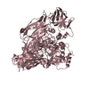
- Assembly
| Deposited unit | 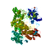
| ||||||||
|---|---|---|---|---|---|---|---|---|---|
| 1 |
| ||||||||
| Unit cell |
|
-Components
| #1: Protein | Mass: 95923.898 Da / Num. of mol.: 1 Source method: isolated from a genetically manipulated source Source: (gene. exp.) Serratia marcescens (bacteria)Description: PERIPLASMATIC TARGETING SEQUENCE RESIDUES 1-27 CLEAVED OFF DURING MATURATION Plasmid: PEMBL18 / Cellular location (production host): PERIPLASM / Production host: | ||||
|---|---|---|---|---|---|
| #2: Chemical | ChemComp-SO4 /   Mass: 96.063 Da / Num. of mol.: 4 / Source method: obtained synthetically / Formula: SO4 Mass: 96.063 Da / Num. of mol.: 4 / Source method: obtained synthetically / Formula: SO4#3: Water | ChemComp-HOH / |  Mass: 18.015 Da / Num. of mol.: 771 / Source method: isolated from a natural source / Formula: H2O Mass: 18.015 Da / Num. of mol.: 771 / Source method: isolated from a natural source / Formula: H2OHas protein modification | Y | |
-Experimental details
-Experiment
| Experiment | Method: X-RAY DIFFRACTION / Number of used crystals: 1 |
|---|
- Sample preparation
| Crystal | Density Matthews: 2.4 Å3/Da / Density % sol: 44.4 % | ||||||||||||||||||||
|---|---|---|---|---|---|---|---|---|---|---|---|---|---|---|---|---|---|---|---|---|---|
| Crystal grow | Method: vapor diffusion, hanging drop / pH: 5.6 Details: CRYSTAL WERE GROWN AT ROOM TEMPERATURE FROM HANGING DROPS CONTAINING 62% SATURATED AMMONIUM SULFATE, 1.5% ISOPROPANOL AND 100 MM CACODYLATE, PH 5.6., vapor diffusion - hanging drop Temp details: room temp | ||||||||||||||||||||
| Crystal | *PLUS | ||||||||||||||||||||
| Crystal grow | *PLUS Method: unknown | ||||||||||||||||||||
| Components of the solutions | *PLUS
|
-Data collection
| Diffraction | Mean temperature: 277 K |
|---|---|
| Diffraction source | Source: SYNCHROTRON / Site: EMBL/DESY, HAMBURG  / Beamline: X11 / Wavelength: 1 / Beamline: X11 / Wavelength: 1 |
| Detector | Type: MARRESEARCH / Detector: IMAGE PLATE / Date: May 10, 1994 / Details: SEGMENTED TOROIDAL MIRROR |
| Radiation | Monochromator: SI(111) / Monochromatic (M) / Laue (L): M / Scattering type: x-ray |
| Radiation wavelength | Wavelength: 1 Å / Relative weight: 1 |
| Reflection | Resolution: 1.85→10 Å / Num. obs: 82854 / % possible obs: 99.9 % / Observed criterion σ(I): -3 / Redundancy: 5.5 % / Biso Wilson estimate: 17.11 Å2 / Rmerge(I) obs: 0.062 / Net I/σ(I): 15.8 |
| Reflection shell | Resolution: 1.85→1.88 Å / Redundancy: 3.2 % / Rmerge(I) obs: 0.276 / Mean I/σ(I) obs: 3.2 / % possible all: 99.9 |
| Reflection | *PLUS Highest resolution: 1.9 Å / Lowest resolution: 15 Å / Num. obs: 83596 |
| Reflection shell | *PLUS % possible obs: 99.8 % |
- Processing
| Software |
| ||||||||||||||||||||||||||||||||||||||||||||||||||||||||||||||||||||||||||||||||||||
|---|---|---|---|---|---|---|---|---|---|---|---|---|---|---|---|---|---|---|---|---|---|---|---|---|---|---|---|---|---|---|---|---|---|---|---|---|---|---|---|---|---|---|---|---|---|---|---|---|---|---|---|---|---|---|---|---|---|---|---|---|---|---|---|---|---|---|---|---|---|---|---|---|---|---|---|---|---|---|---|---|---|---|---|---|---|
| Refinement | Method to determine structure: MIR / Resolution: 1.85→10 Å / Cross valid method: R-FREE / σ(F): -4 Details: RESIDUES 155 AND 156: THERE IS ONLY WEAK DENSITY AND THESE RESIDUES MAY FORM (TWO OR MORE) ALTERNATIVE LOOP CONFORMATIONS.
| ||||||||||||||||||||||||||||||||||||||||||||||||||||||||||||||||||||||||||||||||||||
| Displacement parameters | Biso mean: 17.96 Å2 | ||||||||||||||||||||||||||||||||||||||||||||||||||||||||||||||||||||||||||||||||||||
| Refine analyze | Luzzati d res low obs: 4.5 Å / Luzzati sigma a obs: 0.11 Å | ||||||||||||||||||||||||||||||||||||||||||||||||||||||||||||||||||||||||||||||||||||
| Refinement step | Cycle: LAST / Resolution: 1.85→10 Å
| ||||||||||||||||||||||||||||||||||||||||||||||||||||||||||||||||||||||||||||||||||||
| Refine LS restraints |
| ||||||||||||||||||||||||||||||||||||||||||||||||||||||||||||||||||||||||||||||||||||
| Software | *PLUS Name: PROLSQ / Classification: refinement | ||||||||||||||||||||||||||||||||||||||||||||||||||||||||||||||||||||||||||||||||||||
| Refinement | *PLUS Highest resolution: 1.9 Å / Lowest resolution: 15 Å | ||||||||||||||||||||||||||||||||||||||||||||||||||||||||||||||||||||||||||||||||||||
| Solvent computation | *PLUS | ||||||||||||||||||||||||||||||||||||||||||||||||||||||||||||||||||||||||||||||||||||
| Displacement parameters | *PLUS Biso mean: 17.963 Å2 |
