 Movie
Movie Controller

[English] 日本語
 Yorodumi
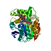
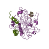
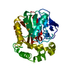
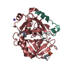
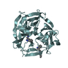
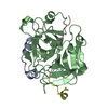
- PDB-1q0r: Crystal structure of aclacinomycin methylesterase (RdmC) with bou... -
+ Open data

- Basic information
| Entry | Database: PDB / ID: 1q0r | ||||||
|---|---|---|---|---|---|---|---|
| Title | Crystal structure of aclacinomycin methylesterase (RdmC) with bound product analogue, 10-decarboxymethylaclacinomycin T (DcmaT) | ||||||
 Components Components | aclacinomycin methylesterase | ||||||
 Keywords Keywords | HYDROLASE / Anthracycline / methylesterase / polyketide / Streptomyces / tailoring enzyme / Structural Proteomics in Europe / SPINE / Structural Genomics | ||||||
| Function / homology |  Function and homology information Function and homology informationaclacinomycin methylesterase / aclacinomycin T methylesterase activity / glycerolipid catabolic process / triacylglycerol lipase activity / antibiotic biosynthetic process Similarity search - Function | ||||||
| Biological species |  Streptomyces purpurascens (bacteria) Streptomyces purpurascens (bacteria) | ||||||
| Method |  X-RAY DIFFRACTION / SYNCHROTRON / MIR / Resolution: 1.45 Å X-RAY DIFFRACTION / SYNCHROTRON / MIR / Resolution: 1.45 Å | ||||||
 Authors Authors | Jansson, A. / Niemi, J. / Mantsala, P. / Schneider, G. / Structural Proteomics in Europe (SPINE) | ||||||
 Citation Citation | Journal: J.Biol.Chem. / Year: 2003 Title: Crystal structure of aclacinomycin methylesterase with bound product analogues: implications for anthracycline recognition and mechanism. Authors: Jansson, A. / Niemi, J. / Mantsala, P. / Schneider, G. #1: Journal: Acta Crystallogr.,Sect.D / Year: 2003Title: Crystallization and preliminary x-ray diffraction studies of aclacinomycin-10-methyl esterase and aclacinomycin-10-hydroxylase from Streptomyces purpurascens Authors: Jansson, A. / Niemi, J. / Mantsala, P. / Schneider, G. | ||||||
| History |
|
- Structure visualization
| Structure viewer | Molecule: MolmilJmol/JSmol |
|---|
- Downloads & links
-Download
| PDBx/mmCIF format | 1q0r.cif.gz | 76.8 KB | Display | PDBx/mmCIF format |
|---|---|---|---|---|
| PDB format | pdb1q0r.ent.gz | 55.6 KB | Display | PDB format |
| PDBx/mmJSON format | 1q0r.json.gz | Tree view | PDBx/mmJSON format | |
| Others |  Other downloads Other downloads |
-Validation report
| Arichive directory | https://data.pdbj.org/pub/pdb/validation_reports/q0/1q0rftp://data.pdbj.org/pub/pdb/validation_reports/q0/1q0r | HTTPS FTP |
|---|
-Related structure data


-Links
 PDBj
PDBj


- Assembly
| Deposited unit | 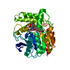
| ||||||||
|---|---|---|---|---|---|---|---|---|---|
| 1 |
| ||||||||
| Unit cell |
|
-Components
| #1: Protein | Mass: 31828.873 Da / Num. of mol.: 1 Source method: isolated from a genetically manipulated source Source: (gene. exp.) Streptomyces purpurascens (bacteria) / Gene: rdmc / Plasmid: pRDM*12 / Species (production host): Streptomyces lividans / Production host: Streptomyces lividans TK24 (bacteria) / Strain (production host): TK24 / References: UniProt: Q54528 |
|---|---|
| #2: Chemical | ChemComp-AKT /   Mass: 511.564 Da / Num. of mol.: 1 / Source method: obtained synthetically / Formula: C28H33NO8 Mass: 511.564 Da / Num. of mol.: 1 / Source method: obtained synthetically / Formula: C28H33NO8 |
| #3: Chemical | ChemComp-SO4 /   Mass: 96.063 Da / Num. of mol.: 1 / Source method: obtained synthetically / Formula: SO4 Mass: 96.063 Da / Num. of mol.: 1 / Source method: obtained synthetically / Formula: SO4 |
| #4: Chemical | ChemComp-1PE /   Mass: 238.278 Da / Num. of mol.: 1 / Source method: obtained synthetically / Formula: C10H22O6 / Comment: precipitant*YM Mass: 238.278 Da / Num. of mol.: 1 / Source method: obtained synthetically / Formula: C10H22O6 / Comment: precipitant*YM |
| #5: Water | ChemComp-HOH /  Mass: 18.015 Da / Num. of mol.: 256 / Source method: isolated from a natural source / Formula: H2O Mass: 18.015 Da / Num. of mol.: 256 / Source method: isolated from a natural source / Formula: H2O |
-Experimental details
-Experiment
| Experiment | Method: X-RAY DIFFRACTION / Number of used crystals: 1 |
|---|
- Sample preparation
| Crystal | Density Matthews: 2.22 Å3/Da / Density % sol: 44.49 % | ||||||||||||||||||||||||||||||||||||
|---|---|---|---|---|---|---|---|---|---|---|---|---|---|---|---|---|---|---|---|---|---|---|---|---|---|---|---|---|---|---|---|---|---|---|---|---|---|
| Crystal grow | Temperature: 294 K / Method: vapor diffusion, hanging drop / pH: 7.5 Details: Ammonium sulphate, PEG400, Hepes, pH 7.5, VAPOR DIFFUSION, HANGING DROP, temperature 294K | ||||||||||||||||||||||||||||||||||||
| Crystal grow | *PLUS Method: vapor diffusion, hanging drop | ||||||||||||||||||||||||||||||||||||
| Components of the solutions | *PLUS
|
-Data collection
| Diffraction | Mean temperature: 100 K |
|---|---|
| Diffraction source | Source: SYNCHROTRON / Site: EMBL/DESY, HAMBURG  / Beamline: X11 / Wavelength: 0.8094 Å / Beamline: X11 / Wavelength: 0.8094 Å |
| Detector | Type: MARRESEARCH / Detector: CCD / Date: Apr 25, 2002 |
| Radiation | Protocol: SINGLE WAVELENGTH / Monochromatic (M) / Laue (L): M / Scattering type: x-ray |
| Radiation wavelength | Wavelength: 0.8094 Å / Relative weight: 1 |
| Reflection | Resolution: 1.45→18.45 Å / Num. all: 34733 / Num. obs: 34733 / % possible obs: 84.8 % / Observed criterion σ(F): 0 / Observed criterion σ(I): 0 / Redundancy: 7.3 % / Biso Wilson estimate: 12 Å2 / Rsym value: 0.041 / Net I/σ(I): 15.1 |
| Reflection shell | Resolution: 1.45→1.53 Å / Mean I/σ(I) obs: 2 / Rsym value: 0.273 / % possible all: 63.5 |
| Reflection | *PLUS Lowest resolution: 20 Å / Num. measured all: 234728 / Rmerge(I) obs: 0.041 |
| Reflection shell | *PLUS % possible obs: 63.5 % / Rmerge(I) obs: 0.273 |
- Processing
| Software |
| ||||||||||||||||||||
|---|---|---|---|---|---|---|---|---|---|---|---|---|---|---|---|---|---|---|---|---|---|
| Refinement | Method to determine structure: MIR / Resolution: 1.45→18.45 Å Isotropic thermal model: Individual isotropic B-factors for each atom Cross valid method: THROUGHOUT / σ(F): 0 / Stereochemistry target values: Maximum likelihood / Details: Riding hydrogens
| ||||||||||||||||||||
| Displacement parameters | Biso mean: 16 Å2 | ||||||||||||||||||||
| Refinement step | Cycle: LAST / Resolution: 1.45→18.45 Å
| ||||||||||||||||||||
| Refine LS restraints |
| ||||||||||||||||||||
| LS refinement shell | Resolution: 1.45→1.51 Å /
| ||||||||||||||||||||
| Refinement | *PLUS % reflection Rfree: 8 % | ||||||||||||||||||||
| Solvent computation | *PLUS | ||||||||||||||||||||
| Displacement parameters | *PLUS | ||||||||||||||||||||
| Refine LS restraints | *PLUS
|
