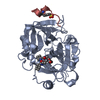
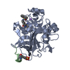
[English] 日本語
Yorodumi
- PDB-3po1: Thrombin in complex with Benzothiazole Guanidine -

+
Open data


ID or keywords:

Loading...

-
Basic information

Entry
Database: PDB / ID: 3po1
TitleThrombin in complex with Benzothiazole Guanidine
Components
  • (Thrombin heavy ...) x 2
  • Thrombin light chain
  • thrombin peptide
KeywordsHYDROLASE/HYDROLASE inhibitor / serine protease / coagulation factor II / HYDROLASE-HYDROLASE inhibitor complex
Function / homology
Function and homology information


positive regulation of lipid kinase activity / positive regulation of phospholipase C-activating G protein-coupled receptor signaling pathway / cytolysis by host of symbiont cells / thrombospondin receptor activity / Defective factor XII causes hereditary angioedema / thrombin / neutrophil-mediated killing of gram-negative bacterium / regulation of blood coagulation / ligand-gated ion channel signaling pathway / Defective F8 cleavage by thrombin ...positive regulation of lipid kinase activity / positive regulation of phospholipase C-activating G protein-coupled receptor signaling pathway / cytolysis by host of symbiont cells / thrombospondin receptor activity / Defective factor XII causes hereditary angioedema / thrombin / neutrophil-mediated killing of gram-negative bacterium / regulation of blood coagulation / ligand-gated ion channel signaling pathway / Defective F8 cleavage by thrombin / Platelet Aggregation (Plug Formation) / negative regulation of platelet activation / negative regulation of astrocyte differentiation / positive regulation of collagen biosynthetic process / negative regulation of cytokine production involved in inflammatory response / positive regulation of blood coagulation / negative regulation of fibrinolysis / Gamma-carboxylation of protein precursors / Transport of gamma-carboxylated protein precursors from the endoplasmic reticulum to the Golgi apparatus / Common Pathway of Fibrin Clot Formation / Removal of aminoterminal propeptides from gamma-carboxylated proteins / fibrinolysis / regulation of cytosolic calcium ion concentration / Intrinsic Pathway of Fibrin Clot Formation / Peptide ligand-binding receptors / positive regulation of release of sequestered calcium ion into cytosol / Regulation of Complement cascade / acute-phase response / Cell surface interactions at the vascular wall / lipopolysaccharide binding / negative regulation of proteolysis / positive regulation of receptor signaling pathway via JAK-STAT / growth factor activity / positive regulation of insulin secretion / platelet activation / response to wounding / Golgi lumen / positive regulation of protein localization to nucleus / Regulation of Insulin-like Growth Factor (IGF) transport and uptake by Insulin-like Growth Factor Binding Proteins (IGFBPs) / positive regulation of reactive oxygen species metabolic process / blood coagulation / antimicrobial humoral immune response mediated by antimicrobial peptide / Thrombin signalling through proteinase activated receptors (PARs) / heparin binding / regulation of cell shape / positive regulation of cell growth / G alpha (q) signalling events / collagen-containing extracellular matrix / blood microparticle / cell surface receptor signaling pathway / positive regulation of phosphatidylinositol 3-kinase/protein kinase B signal transduction / positive regulation of protein phosphorylation / G protein-coupled receptor signaling pathway / endoplasmic reticulum lumen / signaling receptor binding / serine-type endopeptidase activity / calcium ion binding / positive regulation of cell population proliferation / proteolysis / extracellular space / extracellular exosome / extracellular region / plasma membrane
Similarity search - Function
Prothrombin/thrombin / Thrombin light chain / Thrombin light chain domain superfamily / Thrombin light chain / Kringle domain / Kringle / Kringle, conserved site / Kringle superfamily / Kringle domain signature. / Kringle domain profile. ...Prothrombin/thrombin / Thrombin light chain / Thrombin light chain domain superfamily / Thrombin light chain / Kringle domain / Kringle / Kringle, conserved site / Kringle superfamily / Kringle domain signature. / Kringle domain profile. / Kringle domain / Vitamin K-dependent carboxylation/gamma-carboxyglutamic (GLA) domain / Gamma-carboxyglutamic acid-rich (GLA) domain / Gamma-carboxyglutamic acid-rich (GLA) domain superfamily / Vitamin K-dependent carboxylation domain. / Gla domain profile. / Domain containing Gla (gamma-carboxyglutamate) residues. / Kringle-like fold / Serine proteases, trypsin family, histidine active site / Serine proteases, trypsin family, serine active site / Peptidase S1A, chymotrypsin family / Serine proteases, trypsin family, histidine active site. / Serine proteases, trypsin domain profile. / Serine proteases, trypsin family, serine active site. / Trypsin-like serine protease / Serine proteases, trypsin domain / Trypsin / Trypsin-like serine proteases / Thrombin, subunit H / Peptidase S1, PA clan, chymotrypsin-like fold / Peptidase S1, PA clan / Beta Barrel / Mainly Beta
Similarity search - Domain/homology
ACETATE ION / Chem-MKY / Prothrombin
Similarity search - Component
Biological speciesHomo sapiens (human)
MethodX-RAY DIFFRACTION / SYNCHROTRON / MOLECULAR REPLACEMENT / Resolution: 1.65 Å
AuthorsXue, Y.
CitationJournal: Bioorg.Med.Chem.Lett. / Year: 2012
Title: Discovery of benzothiazole guanidines as novel inhibitors of thrombin and trypsin IV.
Authors: Karle, M. / Knecht, W. / Xue, Y.
History
DepositionNov 21, 2010Deposition site: RCSB / Processing site: RCSB
Revision 1.0Nov 23, 2011Provider: repository / Type: Initial release
Revision 1.1Jul 25, 2012Group: Database references

-
Structure visualization

Structure viewerMolecule:
MolmilJmol/JSmol

Downloads & links

-
Assembly

Deposited unit
A: Thrombin light chain
B: Thrombin heavy chain
C: Thrombin heavy chain
D: thrombin peptide
hetero molecules


Theoretical massNumber of molelcules
Total (without water)33,5637
Polymers33,1874
Non-polymers3763
Water2,522140
1


  • Idetical with deposited unit
  • defined by author&software
TypeNameSymmetry operationNumber
identity operation1_555x,y,z1
Buried area10540 Å2
ΔGint-70 kcal/mol
Surface area12250 Å2
MethodPISA
Unit cell
Length a, b, c (Å)68.662, 71.414, 71.880
Angle α, β, γ (deg.)90.000, 99.820, 90.000
Int Tables number5
Space group name H-MC121

-
Components

-
Protein/peptide , 2 types, 2 molecules AD

#1: Protein/peptide Thrombin light chain /


Mass: 3188.627 Da / Num. of mol.: 1 / Source method: isolated from a natural source / Source: (natural) Homo sapiens (human) / References: UniProt: P00734, thrombin
#4: Protein/peptide thrombin peptide


Mass: 1291.337 Da / Num. of mol.: 1 / Source method: isolated from a natural source / Source: (natural) Homo sapiens (human)

-
Thrombin heavy ... , 2 types, 2 molecules BC

#2: Protein Thrombin heavy chain /


Mass: 17070.738 Da / Num. of mol.: 1 / Source method: isolated from a natural source / Source: (natural) Homo sapiens (human) / References: UniProt: P00734, thrombin
#3: Protein Thrombin heavy chain /


Mass: 11636.286 Da / Num. of mol.: 1 / Source method: isolated from a natural source / Source: (natural) Homo sapiens (human) / References: UniProt: P00734, thrombin

-
Non-polymers , 4 types, 143 molecules

#5: Chemical ChemComp-MKY / ethyl [(2Z)-2-(carbamimidoylimino)-6-hydroxy-1,3-benzothiazol-3(2H)-yl]acetate


Mass: 294.330 Da / Num. of mol.: 1 / Source method: obtained synthetically / Formula: C12H14N4O3S
#6: Chemical ChemComp-NA / SODIUM ION


Mass: 22.990 Da / Num. of mol.: 1 / Source method: obtained synthetically / Formula: Na
#7: Chemical ChemComp-ACT / ACETATE ION / Acetate


Mass: 59.044 Da / Num. of mol.: 1 / Source method: obtained synthetically / Formula: C2H3O2
#8: Water ChemComp-HOH / water / Water


Mass: 18.015 Da / Num. of mol.: 140 / Source method: isolated from a natural source / Formula: H2O

-
Experimental details

-
Experiment

ExperimentMethod: X-RAY DIFFRACTION / Number of used crystals: 1

-
Sample preparation

CrystalDensity Matthews: 2.62 Å3/Da / Density % sol: 52.98 %
Crystal growTemperature: 298 K / Method: evaporation / pH: 7.3
Details: 0.05 M sodium phosphate, 28% PEG8000, pH 7.3, EVAPORATION, temperature 298K

-
Data collection

DiffractionMean temperature: 100 K
Diffraction sourceSource: SYNCHROTRON / Site: ESRF / Beamline: ID23-1 / Wavelength: 0.9795 Å
DetectorType: MARMOSAIC 225 mm CCD / Detector: CCD / Date: Sep 15, 2005
RadiationProtocol: SINGLE WAVELENGTH / Monochromatic (M) / Laue (L): M / Scattering type: x-ray
Radiation wavelengthWavelength: 0.9795 Å / Relative weight: 1
ReflectionResolution: 1.65→70.89 Å / Num. all: 39772 / Num. obs: 39772 / % possible obs: 96.8 % / Observed criterion σ(F): 1 / Observed criterion σ(I): 1 / Redundancy: 3.3 % / Biso Wilson estimate: 22.9 Å2 / Rmerge(I) obs: 0.056 / Rsym value: 0.056 / Net I/σ(I): 13.6
Reflection shell

Diffraction-ID: 1

Resolution (Å)Redundancy (%)Rmerge(I) obsMean I/σ(I) obsNum. measured allNum. unique allRsym value% possible all
1.65-1.693.30.342.2925528420.3495.3
1.69-1.743.30.2932.6917328090.29395.4
1.74-1.793.30.2143.5903427580.21495.8
1.79-1.843.30.1734.3887027000.17395.8
1.84-1.913.30.1415.2851625990.14196.4
1.91-1.973.30.1056.8825225130.10596.3
1.97-2.053.30.0847.9801324390.08496.7
2.05-2.133.30.0749.2769723400.07496.9
2.13-2.223.30.0669.7747622750.06697
2.22-2.333.30.0619.8710221590.06197.2
2.33-2.463.30.0599.5685020860.05997.4
2.46-2.613.30.0599.7640019570.05997.7
2.61-2.793.30.0599595818320.05997.9
2.79-3.013.30.0579.8566517320.05798
3.01-3.33.30.0569532316260.05698.3
3.3-3.693.20.0549.8460614190.05498.4
3.69-4.263.20.0511.1414412880.0598.8
4.26-5.223.20.04114.2354410970.04198.8
5.22-7.383.20.04313.927158510.04398.9
7.38-27.1762.90.04113.912974500.04192.6

-
Processing

Software
NameVersionClassificationNB
MOSFLMdata reduction
SCALA3.2.5data scaling
REFMACrefinement
PDB_EXTRACT3.1data extraction
RefinementMethod to determine structure: MOLECULAR REPLACEMENT / Resolution: 1.65→27.176 Å / Cor.coef. Fo:Fc: 0.95 / Cor.coef. Fo:Fc free: 0.928 / WRfactor Rfree: 0.2878 / WRfactor Rwork: 0.2526 / Occupancy max: 1 / Occupancy min: 0.5 / FOM work R set: 0.7908 / SU B: 2.652 / SU ML: 0.089 / SU R Cruickshank DPI: 0.1192 / SU Rfree: 0.1166 / Cross valid method: THROUGHOUT / σ(F): 0 / ESU R Free: 0.117 / Stereochemistry target values: MAXIMUM LIKELIHOOD / Details: HYDROGENS HAVE BEEN ADDED IN THE RIDING POSITIONS
RfactorNum. reflection% reflectionSelection details
Rfree0.2604 1904 4.9 %RANDOM
Rwork0.2271 ---
all0.2287 39772 --
obs0.2287 38491 93.51 %-
Solvent computationIon probe radii: 0.8 Å / Shrinkage radii: 0.8 Å / VDW probe radii: 1.2 Å / Solvent model: BABINET MODEL WITH MASK
Displacement parametersBiso max: 55.38 Å2 / Biso mean: 28.5089 Å2 / Biso min: 13.45 Å2
Baniso -1Baniso -2Baniso -3
1-2.65 Å20 Å20.38 Å2
2---1.35 Å20 Å2
3----1.17 Å2
Refine analyze
FreeObs
Luzzati coordinate error0.117 Å0.119 Å
Refinement stepCycle: LAST / Resolution: 1.65→27.176 Å
ProteinNucleic acidLigandSolventTotal
Num. atoms2327 0 25 140 2492
Refine LS restraints
Refine-IDTypeDev idealDev ideal targetNumber
X-RAY DIFFRACTIONr_bond_refined_d0.0110.0222410
X-RAY DIFFRACTIONr_angle_refined_deg1.3671.9833256
X-RAY DIFFRACTIONr_dihedral_angle_1_deg7.0045282
X-RAY DIFFRACTIONr_dihedral_angle_2_deg33.68723.451113
X-RAY DIFFRACTIONr_dihedral_angle_3_deg13.55615426
X-RAY DIFFRACTIONr_dihedral_angle_4_deg12.5641520
X-RAY DIFFRACTIONr_chiral_restr0.0970.2334
X-RAY DIFFRACTIONr_gen_planes_refined0.0070.021842
X-RAY DIFFRACTIONr_nbd_refined0.2070.21069
X-RAY DIFFRACTIONr_nbtor_refined0.3130.21605
X-RAY DIFFRACTIONr_xyhbond_nbd_refined0.1640.2172
X-RAY DIFFRACTIONr_metal_ion_refined0.0650.24
X-RAY DIFFRACTIONr_symmetry_vdw_refined0.2730.238
X-RAY DIFFRACTIONr_symmetry_hbond_refined0.2190.28
X-RAY DIFFRACTIONr_mcbond_it0.841.51425
X-RAY DIFFRACTIONr_mcangle_it1.47722296
X-RAY DIFFRACTIONr_scbond_it1.95431039
X-RAY DIFFRACTIONr_scangle_it3.0424.5960
LS refinement shellResolution: 1.65→1.693 Å / Total num. of bins used: 20
RfactorNum. reflection% reflection
Rfree0.294 126 -
Rwork0.302 2396 -
all-2522 -
obs--84.38 %

+
About Yorodumi

-
News

-
Feb 9, 2022. New format data for meta-information of EMDB entries

New format data for meta-information of EMDB entries

  • Version 3 of the EMDB header file is now the official format.
  • The previous official version 1.9 will be removed from the archive.

Related info.:EMDB header

External links:wwPDB to switch to version 3 of the EMDB data model

-
Aug 12, 2020. Covid-19 info

Covid-19 info

URL: https://pdbj.org/emnavi/covid19.php

New page: Covid-19 featured information page in EM Navigator.

Related info.:Covid-19 info / Mar 5, 2020. Novel coronavirus structure data

+
Mar 5, 2020. Novel coronavirus structure data

Novel coronavirus structure data

Related info.:Yorodumi Speices / Aug 12, 2020. Covid-19 info

External links:COVID-19 featured content - PDBj / Molecule of the Month (242):Coronavirus Proteases

+
Jan 31, 2019. EMDB accession codes are about to change! (news from PDBe EMDB page)

EMDB accession codes are about to change! (news from PDBe EMDB page)

  • The allocation of 4 digits for EMDB accession codes will soon come to an end. Whilst these codes will remain in use, new EMDB accession codes will include an additional digit and will expand incrementally as the available range of codes is exhausted. The current 4-digit format prefixed with “EMD-” (i.e. EMD-XXXX) will advance to a 5-digit format (i.e. EMD-XXXXX), and so on. It is currently estimated that the 4-digit codes will be depleted around Spring 2019, at which point the 5-digit format will come into force.
  • The EM Navigator/Yorodumi systems omit the EMD- prefix.

Related info.:Q: What is EMD? / ID/Accession-code notation in Yorodumi/EM Navigator

External links:EMDB Accession Codes are Changing Soon! / Contact to PDBj

+
Jul 12, 2017. Major update of PDB

Major update of PDB

  • wwPDB released updated PDB data conforming to the new PDBx/mmCIF dictionary.
  • This is a major update changing the version number from 4 to 5, and with Remediation, in which all the entries are updated.
  • In this update, many items about electron microscopy experimental information are reorganized (e.g. em_software).
  • Now, EM Navigator and Yorodumi are based on the updated data.

External links:wwPDB Remediation / Enriched Model Files Conforming to OneDep Data Standards Now Available in the PDB FTP Archive

-
Yorodumi

Thousand views of thousand structures

  • Yorodumi is a browser for structure data from EMDB, PDB, SASBDB, etc.
  • This page is also the successor to EM Navigator detail page, and also detail information page/front-end page for Omokage search.
  • The word "yorodu" (or yorozu) is an old Japanese word meaning "ten thousand". "mi" (miru) is to see.

Related info.:EMDB / PDB / SASBDB / Comparison of 3 databanks / Yorodumi Search / Aug 31, 2016. New EM Navigator & Yorodumi / Yorodumi Papers / Jmol/JSmol / Function and homology information / Changes in new EM Navigator and Yorodumi

Read more