 Movie
Movie Controller


+ Open data

- Basic information
| Entry | Database: PDB / ID: 1py9 | ||||||
|---|---|---|---|---|---|---|---|
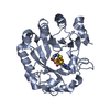
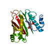
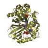
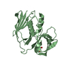
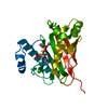
| Title | The crystal structure of an autoantigen in multiple sclerosis | ||||||
 Components Components | Myelin-oligodendrocyte glycoprotein | ||||||
 Keywords Keywords | IMMUNE SYSTEM / myelin sheath / multiple sclerosis / receptor / immunoglobulin / anti-parallel dimer | ||||||
| Function / homology |  Function and homology information Function and homology informationregulation of cytokine production / myelin sheath / T cell receptor signaling pathway / cell adhesion / signaling receptor binding / external side of plasma membrane / cell surface Similarity search - Function | ||||||
| Biological species |  | ||||||
| Method |  X-RAY DIFFRACTION / MOLECULAR REPLACEMENT / Resolution: 1.8 Å X-RAY DIFFRACTION / MOLECULAR REPLACEMENT / Resolution: 1.8 Å | ||||||
 Authors Authors | Clements, C.S. / Reid, H.H. / Beddoe, T. / Tynan, F.E. / Perugini, M.A. / Johns, T.G. / Bernard, C.C. / Rossjohn, J. | ||||||
 Citation Citation | Journal: Proc.Natl.Acad.Sci.USA / Year: 2003 Title: The crystal structure of myelin oligodendrocyte glycoprotein, a key autoantigen in multiple sclerosis Authors: Clements, C.S. / Reid, H.H. / Beddoe, T. / Tynan, F.E. / Perugini, M.A. / Johns, T.G. / Bernard, C.C. / Rossjohn, J. | ||||||
| History |
|
- Structure visualization
| Structure viewer | Molecule: MolmilJmol/JSmol |
|---|
- Downloads & links
-Download
| PDBx/mmCIF format | 1py9.cif.gz | 41.5 KB | Display | PDBx/mmCIF format |
|---|---|---|---|---|
| PDB format | pdb1py9.ent.gz | 28.1 KB | Display | PDB format |
| PDBx/mmJSON format | 1py9.json.gz | Tree view | PDBx/mmJSON format | |
| Others |  Other downloads Other downloads |
-Validation report
| Arichive directory | https://data.pdbj.org/pub/pdb/validation_reports/py/1py9ftp://data.pdbj.org/pub/pdb/validation_reports/py/1py9 | HTTPS FTP |
|---|
-Related structure data
| Similar structure data |
|---|


-Links
 PDBj
PDBj
- Assembly
| Deposited unit | 
| ||||||||
|---|---|---|---|---|---|---|---|---|---|
| 1 | 
| ||||||||
| Unit cell |
| ||||||||
| Components on special symmetry positions |
| ||||||||
| Details | monomer in the asymmtric unit. However a crystallographic, head-to-tail dimer is present |
-Components
| #1: Protein | Mass: 13277.826 Da / Num. of mol.: 1 / Fragment: extracellular domain Source method: isolated from a genetically manipulated source Source: (gene. exp.)  | ||||
|---|---|---|---|---|---|
| #2: Chemical |   Mass: 96.063 Da / Num. of mol.: 2 / Source method: obtained synthetically / Formula: SO4 Mass: 96.063 Da / Num. of mol.: 2 / Source method: obtained synthetically / Formula: SO4#3: Water | ChemComp-HOH / |  Mass: 18.015 Da / Num. of mol.: 215 / Source method: isolated from a natural source / Formula: H2O Mass: 18.015 Da / Num. of mol.: 215 / Source method: isolated from a natural source / Formula: H2OHas protein modification | Y | |
-Experimental details
-Experiment
| Experiment | Method: X-RAY DIFFRACTION / Number of used crystals: 1 |
|---|
- Sample preparation
| Crystal | Density Matthews: 2.25 Å3/Da / Density % sol: 44.87 % | |||||||||||||||||||||||||||||||||||||||||||||||||
|---|---|---|---|---|---|---|---|---|---|---|---|---|---|---|---|---|---|---|---|---|---|---|---|---|---|---|---|---|---|---|---|---|---|---|---|---|---|---|---|---|---|---|---|---|---|---|---|---|---|---|
| Crystal grow | Temperature: 290 K / Method: vapor diffusion, hanging drop / pH: 4.6 Details: PEG, pH 4.6, VAPOR DIFFUSION, HANGING DROP, temperature 290K | |||||||||||||||||||||||||||||||||||||||||||||||||
| Crystal grow | *PLUS pH: 8 / Method: vapor diffusion, hanging drop | |||||||||||||||||||||||||||||||||||||||||||||||||
| Components of the solutions | *PLUS
|
-Data collection
| Diffraction | Mean temperature: 100 K |
|---|---|
| Detector | Type: RIGAKU RAXIS IV / Detector: IMAGE PLATE / Date: Aug 24, 2002 |
| Radiation | Protocol: SINGLE WAVELENGTH / Monochromatic (M) / Laue (L): M / Scattering type: x-ray |
| Radiation wavelength | Relative weight: 1 |
| Reflection | Resolution: 1.8→100 Å / Num. all: 13229 / Num. obs: 13229 / % possible obs: 99 % / Observed criterion σ(F): 0 / Observed criterion σ(I): 0 / Redundancy: 2.5 % / Rmerge(I) obs: 0.065 / Rsym value: 0.069 / Net I/σ(I): 6.9 |
| Reflection shell | Resolution: 1.8→1.9 Å / Rmerge(I) obs: 0.246 / Rsym value: 0.018 / % possible all: 100 |
| Reflection | *PLUS % possible obs: 99 % / Num. measured all: 33022 / Rmerge(I) obs: 0.065 |
| Reflection shell | *PLUS % possible obs: 100 % / Mean I/σ(I) obs: 1.8 |
- Processing
| Software |
| |||||||||||||||||||||||||
|---|---|---|---|---|---|---|---|---|---|---|---|---|---|---|---|---|---|---|---|---|---|---|---|---|---|---|
| Refinement | Method to determine structure: MOLECULAR REPLACEMENT Starting model: B7.2 Resolution: 1.8→100 Å / Cross valid method: THROUGHOUT / σ(F): 0 / Stereochemistry target values: Engh & Huber
| |||||||||||||||||||||||||
| Refinement step | Cycle: LAST / Resolution: 1.8→100 Å
| |||||||||||||||||||||||||
| Refinement | *PLUS Lowest resolution: 50 Å / % reflection Rfree: 3 % | |||||||||||||||||||||||||
| Solvent computation | *PLUS | |||||||||||||||||||||||||
| Displacement parameters | *PLUS | |||||||||||||||||||||||||
| Refine LS restraints | *PLUS
|
