 Movie
Movie Controller

[English] 日本語
 Yorodumi
- PDB-1px2: Crystal Structure of Rat Synapsin I C Domain Complexed to Ca.ATP ... -
+ Open data

- Basic information
| Entry | Database: PDB / ID: 1px2 | ||||||
|---|---|---|---|---|---|---|---|
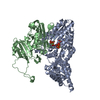
| Title | Crystal Structure of Rat Synapsin I C Domain Complexed to Ca.ATP (Form 1) | ||||||
 Components Components | Synapsin I | ||||||
 Keywords Keywords | MEMBRANE PROTEIN / ATP binding / ATP grasp / calcium (II) ion | ||||||
| Function / homology |  Function and homology information Function and homology informationsynaptic vesicle cycle / synaptic vesicle clustering / Serotonin Neurotransmitter Release Cycle / Dopamine Neurotransmitter Release Cycle / extrinsic component of synaptic vesicle membrane / synaptonemal complex / neurotransmitter secretion / regulation of synaptic vesicle cycle / presynaptic active zone / regulation of synaptic vesicle exocytosis ...synaptic vesicle cycle / synaptic vesicle clustering / Serotonin Neurotransmitter Release Cycle / Dopamine Neurotransmitter Release Cycle / extrinsic component of synaptic vesicle membrane / synaptonemal complex / neurotransmitter secretion / regulation of synaptic vesicle cycle / presynaptic active zone / regulation of synaptic vesicle exocytosis / neuron development / synapse organization / Schaffer collateral - CA1 synapse / terminal bouton / calcium-dependent protein binding / synaptic vesicle / synaptic vesicle membrane / presynapse / cell body / actin binding / cytoskeleton / postsynaptic density / axon / synapse / dendrite / protein kinase binding / Golgi apparatus / ATP binding / identical protein binding Similarity search - Function | ||||||
| Biological species |  | ||||||
| Method |  X-RAY DIFFRACTION / SYNCHROTRON / MOLECULAR REPLACEMENT / Resolution: 2.23 Å X-RAY DIFFRACTION / SYNCHROTRON / MOLECULAR REPLACEMENT / Resolution: 2.23 Å | ||||||
 Authors Authors | Brautigam, C.A. / Chelliah, Y. / Deisenhofer, J. | ||||||
 Citation Citation | Journal: J.Biol.Chem. / Year: 2004 Title: Tetramerization and ATP binding by a protein comprising the A, B, and C domains of rat synapsin I. Authors: Brautigam, C.A. / Chelliah, Y. / Deisenhofer, J. #1: Journal: Embo J. / Year: 1998Title: Synapsin I is structurally similar to ATP-utilizing enzymes Authors: Esser, L. / Wang, C.R. / Hosaka, M. / Smagula, C.S. / Sudhof, T.C. / Deisenhofer, J. | ||||||
| History |
|
- Structure visualization
| Structure viewer | Molecule: MolmilJmol/JSmol |
|---|
- Downloads & links
-Download
| PDBx/mmCIF format | 1px2.cif.gz | 144.6 KB | Display | PDBx/mmCIF format |
|---|---|---|---|---|
| PDB format | pdb1px2.ent.gz | 110.4 KB | Display | PDB format |
| PDBx/mmJSON format | 1px2.json.gz | Tree view | PDBx/mmJSON format | |
| Others |  Other downloads Other downloads |
-Validation report
| Summary document | 1px2_validation.pdf.gz | 1.6 MB | Display | wwPDB validaton report |
|---|---|---|---|---|
| Full document | 1px2_full_validation.pdf.gz | 1.6 MB | Display | |
| Data in XML | 1px2_validation.xml.gz | 27 KB | Display | |
| Data in CIF | 1px2_validation.cif.gz | 38.3 KB | Display | |
| Arichive directory | https://data.pdbj.org/pub/pdb/validation_reports/px/1px2ftp://data.pdbj.org/pub/pdb/validation_reports/px/1px2 | HTTPS FTP |
-Related structure data
| Related structure data |  1pk8C 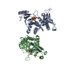 1auxS C: citing same article ( S: Starting model for refinement |
|---|---|
| Similar structure data |


-Links
 PDBj
PDBj


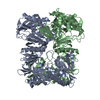
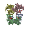
- Assembly
| Deposited unit |
| ||||||||
|---|---|---|---|---|---|---|---|---|---|
| 1 |
| ||||||||
| Unit cell |
| ||||||||
| Details | The second half of the (presumably) biological tetramer is generated by the twofold axis: -y + 1, -x + 1, -z + 1/6 |
-Components
| #1: Protein | Mass: 45539.398 Da / Num. of mol.: 2 / Fragment: A, B, & C domains Source method: isolated from a genetically manipulated source Source: (gene. exp.)  #2: Chemical |   Mass: 40.078 Da / Num. of mol.: 2 / Source method: obtained synthetically / Formula: Ca / Details: Sigma Chemical Corp Mass: 40.078 Da / Num. of mol.: 2 / Source method: obtained synthetically / Formula: Ca / Details: Sigma Chemical Corp#3: Chemical |   Mass: 507.181 Da / Num. of mol.: 2 / Source method: obtained synthetically / Formula: C10H16N5O13P3 / Details: Sigma Chemical Corp / Comment: ATP, energy-carrying molecule*YM Mass: 507.181 Da / Num. of mol.: 2 / Source method: obtained synthetically / Formula: C10H16N5O13P3 / Details: Sigma Chemical Corp / Comment: ATP, energy-carrying molecule*YM#4: Water | ChemComp-HOH / |  Mass: 18.015 Da / Num. of mol.: 326 / Source method: isolated from a natural source / Formula: H2O Mass: 18.015 Da / Num. of mol.: 326 / Source method: isolated from a natural source / Formula: H2O |
|---|
-Experimental details
-Experiment
| Experiment | Method: X-RAY DIFFRACTION / Number of used crystals: 1 |
|---|
- Sample preparation
| Crystal | Density Matthews: 2.23 Å3/Da / Density % sol: 44.89 % | ||||||||||||||||||||||||||||||||||||||||||
|---|---|---|---|---|---|---|---|---|---|---|---|---|---|---|---|---|---|---|---|---|---|---|---|---|---|---|---|---|---|---|---|---|---|---|---|---|---|---|---|---|---|---|---|
| Crystal grow | Temperature: 299 K / Method: vapor diffusion, hanging drop / pH: 7.5 Details: PEGMME 5K, Tris, NaCl, Ca.ATP, EDTA, pH 7.5, VAPOR DIFFUSION, HANGING DROP, temperature 299K | ||||||||||||||||||||||||||||||||||||||||||
| Crystal grow | *PLUS Method: vapor diffusion, hanging drop | ||||||||||||||||||||||||||||||||||||||||||
| Components of the solutions | *PLUS
|
-Data collection
| Diffraction | Mean temperature: 93 K |
|---|---|
| Diffraction source | Source: SYNCHROTRON / Site: CHESS  / Beamline: F1 / Wavelength: 0.942 Å / Beamline: F1 / Wavelength: 0.942 Å |
| Detector | Type: ADSC QUANTUM 4 / Detector: CCD / Date: Jul 2, 2000 |
| Radiation | Monochromator: Horizontally bent Si(111), with monochromatic mirrors of Rh-coated Si Protocol: SINGLE WAVELENGTH / Monochromatic (M) / Laue (L): M / Scattering type: x-ray |
| Radiation wavelength | Wavelength: 0.942 Å / Relative weight: 1 |
| Reflection | Resolution: 2.23→40 Å / Num. all: 41721 / Num. obs: 41721 / % possible obs: 99.8 % / Redundancy: 11.2 % / Biso Wilson estimate: 28.5 Å2 / Rsym value: 0.082 / Net I/σ(I): 29.9 |
| Reflection shell | Resolution: 2.23→2.31 Å / Redundancy: 9 % / Mean I/σ(I) obs: 6.1 / Num. unique all: 4060 / Rsym value: 0.391 / % possible all: 100 |
| Reflection | *PLUS Lowest resolution: 20 Å / Num. obs: 40542 / Num. measured all: 467389 / Rmerge(I) obs: 0.082 |
| Reflection shell | *PLUS % possible obs: 100 % / Rmerge(I) obs: 0.391 |
- Processing
| Software |
| |||||||||||||||||||||||||
|---|---|---|---|---|---|---|---|---|---|---|---|---|---|---|---|---|---|---|---|---|---|---|---|---|---|---|
| Refinement | Method to determine structure: MOLECULAR REPLACEMENT Starting model: PDB entry 1AUX, Ca and ATP-gamma-S removed Resolution: 2.23→20 Å / Rfactor Rfree error: 0.005 / Isotropic thermal model: Restrained / Cross valid method: THROUGHOUT / σ(F): 0 / Stereochemistry target values: modified Engh & Huber Details: Although the A, B, and C domains of rat synapsin I were included in crystallization, only the C domain was observed. Some residues have side chains that are set to occupancies of 0.00 due to disorder.
| |||||||||||||||||||||||||
| Solvent computation | Solvent model: flat model / Bsol: 40.9992 Å2 / ksol: 0.326988 e/Å3 | |||||||||||||||||||||||||
| Displacement parameters | Biso mean: 39.2 Å2
| |||||||||||||||||||||||||
| Refine analyze |
| |||||||||||||||||||||||||
| Refinement step | Cycle: LAST / Resolution: 2.23→20 Å
| |||||||||||||||||||||||||
| Refine LS restraints |
| |||||||||||||||||||||||||
| LS refinement shell | Resolution: 2.23→2.37 Å / Rfactor Rfree error: 0.017
| |||||||||||||||||||||||||
| Refinement | *PLUS % reflection Rfree: 5 % / Rfactor Rfree: 0.24 | |||||||||||||||||||||||||
| Solvent computation | *PLUS | |||||||||||||||||||||||||
| Displacement parameters | *PLUS | |||||||||||||||||||||||||
| Refine LS restraints | *PLUS
|
