 Movie
Movie Controller


+ Open data

- Basic information
| Entry | Database: PDB / ID: 1pk8 | ||||||
|---|---|---|---|---|---|---|---|
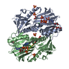
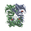
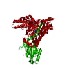
| Title | Crystal Structure of Rat Synapsin I C Domain Complexed to Ca.ATP | ||||||
 Components Components | rat synapsin I | ||||||
 Keywords Keywords | MEMBRANE PROTEIN / ATP binding / ATP grasp / calcium (II) ion | ||||||
| Function / homology |  Function and homology information Function and homology informationsynaptic vesicle cycle / synaptic vesicle clustering / Serotonin Neurotransmitter Release Cycle / Dopamine Neurotransmitter Release Cycle / extrinsic component of synaptic vesicle membrane / synaptonemal complex / neurotransmitter secretion / regulation of synaptic vesicle cycle / presynaptic active zone / regulation of synaptic vesicle exocytosis ...synaptic vesicle cycle / synaptic vesicle clustering / Serotonin Neurotransmitter Release Cycle / Dopamine Neurotransmitter Release Cycle / extrinsic component of synaptic vesicle membrane / synaptonemal complex / neurotransmitter secretion / regulation of synaptic vesicle cycle / presynaptic active zone / regulation of synaptic vesicle exocytosis / neuron development / synapse organization / Schaffer collateral - CA1 synapse / calcium-dependent protein binding / terminal bouton / synaptic vesicle / synaptic vesicle membrane / presynapse / actin binding / cell body / cytoskeleton / postsynaptic density / axon / synapse / dendrite / protein kinase binding / Golgi apparatus / ATP binding / identical protein binding Similarity search - Function | ||||||
| Biological species |  | ||||||
| Method |  X-RAY DIFFRACTION / SYNCHROTRON / MOLECULAR REPLACEMENT / Resolution: 2.1 Å X-RAY DIFFRACTION / SYNCHROTRON / MOLECULAR REPLACEMENT / Resolution: 2.1 Å | ||||||
 Authors Authors | Brautigam, C.A. / Chelliah, Y. / Deisenhofer, J. | ||||||
 Citation Citation | Journal: J.Biol.Chem. / Year: 2004 Title: Tetramerization and ATP binding by a protein comprising the A, B, and C domains of rat synapsin I. Authors: Brautigam, C.A. / Chelliah, Y. / Deisenhofer, J. #1: Journal: Embo J. / Year: 1998Title: Synapsin I is structurally similar to ATP-utilizing enzymes Authors: Esser, L. / Wang, C.R. / Hosaka, M. / Smagula, C.S. / Sudhof, T.C. / Deisenhofer, J. | ||||||
| History |
|
- Structure visualization
| Structure viewer | Molecule: MolmilJmol/JSmol |
|---|
- Downloads & links
-Download
| PDBx/mmCIF format | 1pk8.cif.gz | 501.1 KB | Display | PDBx/mmCIF format |
|---|---|---|---|---|
| PDB format | pdb1pk8.ent.gz | 404.2 KB | Display | PDB format |
| PDBx/mmJSON format | 1pk8.json.gz | Tree view | PDBx/mmJSON format | |
| Others |  Other downloads Other downloads |
-Validation report
| Arichive directory | https://data.pdbj.org/pub/pdb/validation_reports/pk/1pk8ftp://data.pdbj.org/pub/pdb/validation_reports/pk/1pk8 | HTTPS FTP |
|---|
-Related structure data
| Related structure data | 1px2C 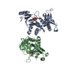 1auxS S: Starting model for refinement C: citing same article ( |
|---|---|
| Similar structure data |


-Links
 PDBj
PDBj


- Assembly
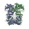
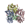
| Deposited unit | 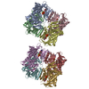
| ||||||||
|---|---|---|---|---|---|---|---|---|---|
| 1 | 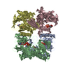
| ||||||||
| 2 | 
| ||||||||
| Unit cell |
|
-Components
| #1: Protein | Mass: 45539.398 Da / Num. of mol.: 8 / Fragment: A, B & C domains Source method: isolated from a genetically manipulated source Source: (gene. exp.)  #2: Chemical | ChemComp-CA /   Mass: 40.078 Da / Num. of mol.: 8 / Source method: obtained synthetically / Formula: Ca / Details: Sigma Chemical Corp. Mass: 40.078 Da / Num. of mol.: 8 / Source method: obtained synthetically / Formula: Ca / Details: Sigma Chemical Corp.#3: Chemical | ChemComp-ATP /   Mass: 507.181 Da / Num. of mol.: 8 / Source method: obtained synthetically / Formula: C10H16N5O13P3 / Details: Sigma Chemical Corp. / Comment: ATP, energy-carrying molecule*YM Mass: 507.181 Da / Num. of mol.: 8 / Source method: obtained synthetically / Formula: C10H16N5O13P3 / Details: Sigma Chemical Corp. / Comment: ATP, energy-carrying molecule*YM#4: Chemical |   Mass: 62.068 Da / Num. of mol.: 2 / Source method: obtained synthetically / Formula: C2H6O2 Mass: 62.068 Da / Num. of mol.: 2 / Source method: obtained synthetically / Formula: C2H6O2#5: Water | ChemComp-HOH / |  Mass: 18.015 Da / Num. of mol.: 665 / Source method: isolated from a natural source / Formula: H2O / Details: Sigma Chemical Corp. Mass: 18.015 Da / Num. of mol.: 665 / Source method: isolated from a natural source / Formula: H2O / Details: Sigma Chemical Corp. |
|---|
-Experimental details
-Experiment
| Experiment | Method: X-RAY DIFFRACTION / Number of used crystals: 1 |
|---|
- Sample preparation
| Crystal | Density Matthews: 1.89 Å3/Da / Density % sol: 34.82 % | ||||||||||||||||||||||||||||||||||||||||||
|---|---|---|---|---|---|---|---|---|---|---|---|---|---|---|---|---|---|---|---|---|---|---|---|---|---|---|---|---|---|---|---|---|---|---|---|---|---|---|---|---|---|---|---|
| Crystal grow | Temperature: 299 K / Method: vapor diffusion, hanging drop / pH: 7.5 Details: PEGMME 5000, Tris, NaCl, Ca.ATP, EDTA, DTT, pH 7.5, VAPOR DIFFUSION, HANGING DROP, temperature 299K | ||||||||||||||||||||||||||||||||||||||||||
| Crystal grow | *PLUS Method: vapor diffusion, hanging drop | ||||||||||||||||||||||||||||||||||||||||||
| Components of the solutions | *PLUS
|
-Data collection
| Diffraction | Mean temperature: 100 K |
|---|---|
| Diffraction source | Source: SYNCHROTRON / Site: APS  / Beamline: 19-ID / Wavelength: 1.008 Å / Beamline: 19-ID / Wavelength: 1.008 Å |
| Detector | Type: CUSTOM-MADE / Detector: CCD / Date: Mar 17, 2000 |
| Radiation | Monochromator: Double crystal monochromator, Si-111 / Protocol: SINGLE WAVELENGTH / Monochromatic (M) / Laue (L): M / Scattering type: x-ray |
| Radiation wavelength | Wavelength: 1.008 Å / Relative weight: 1 |
| Reflection | Resolution: 2.1→40 Å / Num. obs: 150398 / % possible obs: 96.3 % / Redundancy: 3.7 % / Biso Wilson estimate: 19 Å2 / Rsym value: 0.058 / Net I/σ(I): 20.9 |
| Reflection shell | Resolution: 2.1→2.18 Å / Redundancy: 3.6 % / Mean I/σ(I) obs: 2.8 / Num. unique all: 14381 / Rsym value: 0.373 / % possible all: 92 |
| Reflection | *PLUS Lowest resolution: 20 Å / Num. obs: 144586 / Num. measured all: 522852 / Rmerge(I) obs: 0.058 |
| Reflection shell | *PLUS % possible obs: 92 % / Rmerge(I) obs: 0.373 |
- Processing
| Software |
| |||||||||||||||||||||||||
|---|---|---|---|---|---|---|---|---|---|---|---|---|---|---|---|---|---|---|---|---|---|---|---|---|---|---|
| Refinement | Method to determine structure: MOLECULAR REPLACEMENT Starting model: PDB ENTRY 1AUX, ATPgammaS and Ca2+ removed Resolution: 2.1→20 Å / Isotropic thermal model: restrained / Cross valid method: THROUGHOUT / σ(F): 0 / Stereochemistry target values: MODIFIED ENGH & HUBER Details: Although the A, B, and C domains of rat Synapsin I were included in crystallization, only the C domain was observed. Some side chain atoms were set to an occupancy of 0.00 because of ...Details: Although the A, B, and C domains of rat Synapsin I were included in crystallization, only the C domain was observed. Some side chain atoms were set to an occupancy of 0.00 because of disorder. Two conformations of ATP were modeled and refined due to ample evidence in the electron density maps. The differing phosphate positions would probably necessitate a movement in the associated Ca2+ ions, but only one Ca2+ was modeled per pair of ATP conformations
| |||||||||||||||||||||||||
| Solvent computation | Solvent model: flat model / Bsol: 37.4817 Å2 / ksol: 0.341215 e/Å3 | |||||||||||||||||||||||||
| Displacement parameters | Biso mean: 34.2 Å2
| |||||||||||||||||||||||||
| Refine analyze |
| |||||||||||||||||||||||||
| Refinement step | Cycle: LAST / Resolution: 2.1→20 Å
| |||||||||||||||||||||||||
| Refine LS restraints |
| |||||||||||||||||||||||||
| LS refinement shell | Resolution: 2.1→2.23 Å / Rfactor Rfree error: 0.007
| |||||||||||||||||||||||||
| Refinement | *PLUS % reflection Rfree: 10 % | |||||||||||||||||||||||||
| Solvent computation | *PLUS | |||||||||||||||||||||||||
| Displacement parameters | *PLUS | |||||||||||||||||||||||||
| Refine LS restraints | *PLUS
|
