 Movie
Movie Controller

[English] 日本語
 Yorodumi
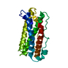
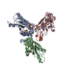
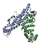
- PDB-1pvh: Crystal structure of leukemia inhibitory factor in complex with gp130 -
+ Open data

- Basic information
| Entry | Database: PDB / ID: 1pvh | ||||||
|---|---|---|---|---|---|---|---|
| Title | Crystal structure of leukemia inhibitory factor in complex with gp130 | ||||||
 Components Components |
| ||||||
 Keywords Keywords | signaling protein/cytokine / cytokine / receptor / signaling / beta sheet / four helix bundle / signaling protein-cytokine COMPLEX | ||||||
| Function / homology |  Function and homology information Function and homology informationleukemia inhibitory factor receptor binding / positive regulation of mesenchymal to epithelial transition involved in metanephros morphogenesis / meiotic nuclear division / negative regulation of meiotic nuclear division / muscle organ morphogenesis / type I oncostatin-M receptor complex / interleukin-27 receptor activity / oncostatin-M-mediated signaling pathway / ciliary neurotrophic factor receptor activity / ciliary neurotrophic factor receptor binding ...leukemia inhibitory factor receptor binding / positive regulation of mesenchymal to epithelial transition involved in metanephros morphogenesis / meiotic nuclear division / negative regulation of meiotic nuclear division / muscle organ morphogenesis / type I oncostatin-M receptor complex / interleukin-27 receptor activity / oncostatin-M-mediated signaling pathway / ciliary neurotrophic factor receptor activity / ciliary neurotrophic factor receptor binding / leukemia inhibitory factor signaling pathway / interleukin-11 receptor activity / interleukin-11 binding / interleukin-6 receptor activity / negative regulation of interleukin-6-mediated signaling pathway / ciliary neurotrophic factor receptor complex / regulation of metanephric nephron tubule epithelial cell differentiation / ciliary neurotrophic factor-mediated signaling pathway / interleukin-6 receptor complex / interleukin-27-mediated signaling pathway / negative regulation of hormone secretion / trophoblast giant cell differentiation / interleukin-11-mediated signaling pathway / positive regulation of receptor signaling pathway via STAT / T-helper 17 cell lineage commitment / lung vasculature development / positive regulation of acute inflammatory response / positive regulation of astrocyte differentiation / lung lobe morphogenesis / positive regulation of macrophage differentiation / positive regulation of adaptive immune response / intestinal epithelial cell development / positive regulation of peptidyl-tyrosine phosphorylation / positive regulation of platelet aggregation / IL-6-type cytokine receptor ligand interactions / Interleukin-27 signaling / Interleukin-35 Signalling / cell surface receptor signaling pathway via STAT / cytokine receptor activity / positive regulation of cell adhesion mediated by integrin / Interleukin-6 signaling / lung alveolus development / glycogen metabolic process / interleukin-6-mediated signaling pathway / growth factor binding / positive regulation of Notch signaling pathway / MAPK3 (ERK1) activation / positive regulation of cardiac muscle hypertrophy / MAPK1 (ERK2) activation / cytokine binding / Interleukin-10 signaling / decidualization / regulation of cell differentiation / protein tyrosine kinase activator activity / macrophage differentiation / somatic stem cell population maintenance / positive regulation of peptidyl-serine phosphorylation / positive regulation of vascular endothelial growth factor production / positive regulation of osteoblast differentiation / blood vessel remodeling / neuron development / coreceptor activity / response to cytokine / embryo implantation / positive regulation of T cell proliferation / cytokine activity / stem cell differentiation / growth factor activity / negative regulation of ERK1 and ERK2 cascade / positive regulation of fibroblast proliferation / cell morphogenesis / cytokine-mediated signaling pathway / Interleukin-4 and Interleukin-13 signaling / fibroblast proliferation / scaffold protein binding / gene expression / negative regulation of neuron apoptotic process / response to hypoxia / positive regulation of MAPK cascade / signaling receptor complex / immune response / membrane raft / signaling receptor binding / external side of plasma membrane / negative regulation of cell population proliferation / neuronal cell body / positive regulation of cell population proliferation / positive regulation of gene expression / dendrite / negative regulation of apoptotic process / positive regulation of transcription by RNA polymerase II / : / extracellular exosome / extracellular region / membrane / identical protein binding / plasma membrane / cytosol Similarity search - Function | ||||||
| Biological species |  Homo sapiens (human) Homo sapiens (human) | ||||||
| Method |  X-RAY DIFFRACTION / SYNCHROTRON / MOLECULAR REPLACEMENT / Resolution: 2.5 Å X-RAY DIFFRACTION / SYNCHROTRON / MOLECULAR REPLACEMENT / Resolution: 2.5 Å | ||||||
 Authors Authors | Boulanger, M.J. / Bankovich, A.J. / Kortemme, T. / Baker, D. / Garcia, K.C. | ||||||
 Citation Citation | Journal: Mol.Cell / Year: 2003 Title: Convergent mechanisms for recognition of divergent cytokines by the shared signaling receptor gp130. Authors: Boulanger, M.J. / Bankovich, A.J. / Kortemme, T. / Baker, D. / Garcia, K.C. | ||||||
| History |
|
- Structure visualization
| Structure viewer | Molecule: MolmilJmol/JSmol |
|---|
- Downloads & links
-Download
| PDBx/mmCIF format | 1pvh.cif.gz | 157.5 KB | Display | PDBx/mmCIF format |
|---|---|---|---|---|
| PDB format | pdb1pvh.ent.gz | 125.9 KB | Display | PDB format |
| PDBx/mmJSON format | 1pvh.json.gz | Tree view | PDBx/mmJSON format | |
| Others |  Other downloads Other downloads |
-Validation report
| Arichive directory | https://data.pdbj.org/pub/pdb/validation_reports/pv/1pvhftp://data.pdbj.org/pub/pdb/validation_reports/pv/1pvh | HTTPS FTP |
|---|
-Related structure data


-Links
 PDBj
PDBj


- Assembly
| Deposited unit | 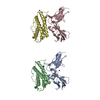
| ||||||||
|---|---|---|---|---|---|---|---|---|---|
| 1 | 
| ||||||||
| 2 | 
| ||||||||
| Unit cell |
|
-Components
| #1: Protein | Mass: 23069.916 Da / Num. of mol.: 2 / Fragment: domains D2 and D3 Source method: isolated from a genetically manipulated source Source: (gene. exp.) Homo sapiens (human) / Gene: IL6ST / Production host:   Spodoptera frugiperda (fall armyworm) / References: UniProt: P40189 Spodoptera frugiperda (fall armyworm) / References: UniProt: P40189#2: Protein | Mass: 18648.596 Da / Num. of mol.: 2 Source method: isolated from a genetically manipulated source Source: (gene. exp.) Homo sapiens (human) / Gene: LIF OR HILDA / Production host: Spodoptera frugiperda (fall armyworm) / References: UniProt: P15018#3: Chemical |   Mass: 126.904 Da / Num. of mol.: 2 / Source method: obtained synthetically / Formula: I Mass: 126.904 Da / Num. of mol.: 2 / Source method: obtained synthetically / Formula: I#4: Water | ChemComp-HOH / |  Mass: 18.015 Da / Num. of mol.: 192 / Source method: isolated from a natural source / Formula: H2O Mass: 18.015 Da / Num. of mol.: 192 / Source method: isolated from a natural source / Formula: H2OHas protein modification | Y | |
|---|
-Experimental details
-Experiment
| Experiment | Method: X-RAY DIFFRACTION / Number of used crystals: 1 |
|---|
- Sample preparation
| Crystal | Density Matthews: 3.03 Å3/Da / Density % sol: 59.43 % | ||||||||||||||||||||||||||||||
|---|---|---|---|---|---|---|---|---|---|---|---|---|---|---|---|---|---|---|---|---|---|---|---|---|---|---|---|---|---|---|---|
| Crystal grow | Temperature: 298 K / Method: vapor diffusion, sitting drop / pH: 7.5 Details: PEG 3350, Sodium iodide, Imidazole, pH 7.5, VAPOR DIFFUSION, SITTING DROP, temperature 298K | ||||||||||||||||||||||||||||||
| Crystal grow | *PLUS Method: vapor diffusion, sitting drop | ||||||||||||||||||||||||||||||
| Components of the solutions | *PLUS
|
-Data collection
| Diffraction | Mean temperature: 100 K |
|---|---|
| Diffraction source | Source: SYNCHROTRON / Site: ALS  / Beamline: 8.2.1 / Wavelength: 1 Å / Beamline: 8.2.1 / Wavelength: 1 Å |
| Detector | Type: ADSC QUANTUM 210 / Detector: CCD / Date: Aug 15, 2002 |
| Radiation | Protocol: SINGLE WAVELENGTH / Monochromatic (M) / Laue (L): M / Scattering type: x-ray |
| Radiation wavelength | Wavelength: 1 Å / Relative weight: 1 |
| Reflection | Resolution: 2.5→50 Å / Num. all: 35908 / Num. obs: 32573 / % possible obs: 91 % / Observed criterion σ(F): 2 / Observed criterion σ(I): 1 / Biso Wilson estimate: 46.1 Å2 / Rmerge(I) obs: 0.085 / Net I/σ(I): 10.4 |
| Reflection shell | Resolution: 2.5→2.63 Å / Rmerge(I) obs: 0.511 / Mean I/σ(I) obs: 1.5 / % possible all: 78.4 |
| Reflection | *PLUS Num. obs: 35908 / % possible obs: 91 % / Num. measured all: 463823 |
| Reflection shell | *PLUS % possible obs: 78.4 % |
- Processing
| Software |
| ||||||||||||||||||||||||||||||||||||
|---|---|---|---|---|---|---|---|---|---|---|---|---|---|---|---|---|---|---|---|---|---|---|---|---|---|---|---|---|---|---|---|---|---|---|---|---|---|
| Refinement | Method to determine structure: MOLECULAR REPLACEMENT Starting model: LIF - PDB ENTRY 1LKI, GP130 - PDB ENTRY 1I1R Resolution: 2.5→39.86 Å / Rfactor Rfree error: 0.006 / Isotropic thermal model: RESTRAINED / Cross valid method: THROUGHOUT / σ(F): 0 / Stereochemistry target values: Engh & Huber
| ||||||||||||||||||||||||||||||||||||
| Solvent computation | Solvent model: FLAT MODEL / Bsol: 28.8599 Å2 / ksol: 0.316798 e/Å3 | ||||||||||||||||||||||||||||||||||||
| Displacement parameters | Biso mean: 53.3 Å2
| ||||||||||||||||||||||||||||||||||||
| Refine analyze |
| ||||||||||||||||||||||||||||||||||||
| Refinement step | Cycle: LAST / Resolution: 2.5→39.86 Å
| ||||||||||||||||||||||||||||||||||||
| Refine LS restraints |
| ||||||||||||||||||||||||||||||||||||
| LS refinement shell | Resolution: 2.5→2.66 Å / Rfactor Rfree error: 0.021 / Total num. of bins used: 6
| ||||||||||||||||||||||||||||||||||||
| Xplor file |
| ||||||||||||||||||||||||||||||||||||
| Refinement | *PLUS Highest resolution: 2.5 Å / Lowest resolution: 40 Å / % reflection Rfree: 5 % / Rfactor Rfree: 0.288 | ||||||||||||||||||||||||||||||||||||
| Solvent computation | *PLUS | ||||||||||||||||||||||||||||||||||||
| Displacement parameters | *PLUS | ||||||||||||||||||||||||||||||||||||
| Refine LS restraints | *PLUS
| ||||||||||||||||||||||||||||||||||||
| LS refinement shell | *PLUS Rfactor Rfree: 0.415 / Rfactor Rwork: 0.374 |
