 Movie
Movie Controller

[English] 日本語
 Yorodumi
- PDB-1ppf: X-RAY CRYSTAL STRUCTURE OF THE COMPLEX OF HUMAN LEUKOCYTE ELASTAS... -
+ Open data

- Basic information
| Entry | Database: PDB / ID: 1ppf | |||||||||
|---|---|---|---|---|---|---|---|---|---|---|
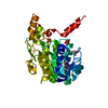
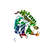
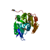
| Title | X-RAY CRYSTAL STRUCTURE OF THE COMPLEX OF HUMAN LEUKOCYTE ELASTASE (PMN ELASTASE) AND THE THIRD DOMAIN OF THE TURKEY OVOMUCOID INHIBITOR | |||||||||
 Components Components |
| |||||||||
 Keywords Keywords | HYDROLASE/HYDROLASE INHIBITOR / SERINE PROTEINASE / HYDROLASE-HYDROLASE INHIBITOR COMPLEX | |||||||||
| Function / homology |  Function and homology information Function and homology informationleukocyte elastase / biosynthetic process of antibacterial peptides active against Gram-negative bacteria / Expression of NOTCH2NL genes / acute inflammatory response to antigenic stimulus / neutrophil-mediated killing of fungus / negative regulation of chemotaxis / positive regulation of leukocyte tethering or rolling / leukocyte migration involved in inflammatory response / response to yeast / negative regulation of chemokine production ...leukocyte elastase / biosynthetic process of antibacterial peptides active against Gram-negative bacteria / Expression of NOTCH2NL genes / acute inflammatory response to antigenic stimulus / neutrophil-mediated killing of fungus / negative regulation of chemotaxis / positive regulation of leukocyte tethering or rolling / leukocyte migration involved in inflammatory response / response to yeast / negative regulation of chemokine production / negative regulation of interleukin-8 production / Antimicrobial peptides / molecular function inhibitor activity / neutrophil-mediated killing of gram-negative bacterium / Activation of Matrix Metalloproteinases / positive regulation of MAP kinase activity / cytokine binding / Collagen degradation / extracellular matrix disassembly / pyroptotic inflammatory response / Pyroptosis / response to UV / phagocytosis / Degradation of the extracellular matrix / phagocytic vesicle / transcription repressor complex / secretory granule / positive regulation of smooth muscle cell proliferation / Regulation of Complement cascade / positive regulation of interleukin-8 production / protein catabolic process / serine-type endopeptidase inhibitor activity / negative regulation of inflammatory response / specific granule lumen / positive regulation of immune response / intracellular calcium ion homeostasis / azurophil granule lumen / transcription corepressor activity / peptidase activity / heparin binding / extracellular matrix / protease binding / endopeptidase activity / response to lipopolysaccharide / defense response to bacterium / serine-type endopeptidase activity / Neutrophil degranulation / cell surface / negative regulation of transcription by RNA polymerase II / Golgi apparatus / proteolysis / : / extracellular exosome / extracellular region / cytoplasm / cytosol Similarity search - Function | |||||||||
| Biological species |  Homo sapiens (human) Homo sapiens (human) | |||||||||
| Method |  X-RAY DIFFRACTION / Resolution: 1.8 Å X-RAY DIFFRACTION / Resolution: 1.8 Å | |||||||||
 Authors Authors | Bode, W. / Wei, A-Z. | |||||||||
 Citation Citation | Journal: EMBO J. / Year: 1986 Title: X-ray crystal structure of the complex of human leukocyte elastase (PMN elastase) and the third domain of the turkey ovomucoid inhibitor. Authors: Bode, W. / Wei, A.Z. / Huber, R. / Meyer, E. / Travis, J. / Neumann, S. #2: Journal: Biochemistry / Year: 1989Title: Human Leukocyte and Porcine Pancreatic Elastase: X-Ray Crystal Structure, Mechanism, Substrate Specificity, and Mechanism-Based Inhibitors Authors: Bode, W. / Meyer, E. / Powers, J.C. #3: Journal: FEBS Lett. / Year: 1988Title: The Refined 2.3 Angstroms Crystal Structure of Human Leukocyte Elastase in a Complex with a Valine Chloromethyl Ketone Inhibitor Authors: Wei, A.Z. / Mayr, I. / Bode, W. #4: Journal: Proc.Natl.Acad.Sci.USA / Year: 1987Title: Primary Structure of Human Neutrophil Elastase Authors: Sinha, S. / Watorek, W. / Karr, S. / Giles, J. / Bode, W. / Travis, J. | |||||||||
| History |
| |||||||||
| Remark 700 | SHEET THE SHEETS PRESENTED AS *B1* AND *B2* ON SHEET RECORDS BELOW ARE ACTUALLY SIX-STRANDED BETA- ...SHEET THE SHEETS PRESENTED AS *B1* AND *B2* ON SHEET RECORDS BELOW ARE ACTUALLY SIX-STRANDED BETA-BARRELS. THESE ARE REPRESENTED BY SEVEN-STRANDED SHEETS IN WHICH THE FIRST AND LAST STRANDS ARE IDENTICAL. |
- Structure visualization
| Structure viewer | Molecule: MolmilJmol/JSmol |
|---|
- Downloads & links
-Download
| PDBx/mmCIF format | 1ppf.cif.gz | 80.4 KB | Display | PDBx/mmCIF format |
|---|---|---|---|---|
| PDB format | pdb1ppf.ent.gz | 58.4 KB | Display | PDB format |
| PDBx/mmJSON format | 1ppf.json.gz | Tree view | PDBx/mmJSON format | |
| Others |  Other downloads Other downloads |
-Validation report
| Arichive directory | https://data.pdbj.org/pub/pdb/validation_reports/pp/1ppfftp://data.pdbj.org/pub/pdb/validation_reports/pp/1ppf | HTTPS FTP |
|---|
-Related structure data
| Similar structure data |
|---|


-Links
 PDBj
PDBj


- Assembly
| Deposited unit | 
| ||||||||
|---|---|---|---|---|---|---|---|---|---|
| 1 |
| ||||||||
| Unit cell |
| ||||||||
| Atom site foot note | 1: RESIDUE PRO I 12 IS A CIS PROLINE. |
-Components
| #1: Protein | Mass: 23318.982 Da / Num. of mol.: 1 Source method: isolated from a genetically manipulated source Source: (gene. exp.) Homo sapiens (human) / References: UniProt: P08246, leukocyte elastase |
|---|---|
| #2: Protein | Mass: 6026.811 Da / Num. of mol.: 1 Source method: isolated from a genetically manipulated source Source: (gene. exp.) |
| #3: Polysaccharide | beta-D-galactopyranose-(1-4)-2-acetamido-2-deoxy-beta-D-glucopyranose-(1-2)-alpha-D-mannopyranose- ...beta-D-galactopyranose-(1-4)-2-acetamido-2-deoxy-beta-D-glucopyranose-(1-2)-alpha-D-mannopyranose-(1-6)-[alpha-D-mannopyranose-(1-3)]beta-D-mannopyranose-(1-4)-2-acetamido-2-deoxy-beta-D-glucopyranose-(1-4)-[alpha-L-fucopyranose-(1-6)]2-acetamido-2-deoxy-beta-D-glucopyranose Source method: isolated from a genetically manipulated source |
| #4: Polysaccharide | alpha-D-glucopyranose-(1-4)-2-acetamido-2-deoxy-beta-D-glucopyranose-(1-2)-alpha-D-mannopyranose-(1- ...alpha-D-glucopyranose-(1-4)-2-acetamido-2-deoxy-beta-D-glucopyranose-(1-2)-alpha-D-mannopyranose-(1-6)-[alpha-D-mannopyranose-(1-3)]beta-D-mannopyranose-(1-4)-2-acetamido-2-deoxy-beta-D-glucopyranose-(1-4)-[alpha-L-fucopyranose-(1-6)]2-acetamido-2-deoxy-beta-D-glucopyranose Source method: isolated from a genetically manipulated source |
| #5: Water | ChemComp-HOH /  Mass: 18.015 Da / Num. of mol.: 272 / Source method: isolated from a natural source / Formula: H2O Mass: 18.015 Da / Num. of mol.: 272 / Source method: isolated from a natural source / Formula: H2O |
| Has protein modification | Y |
| Nonpolymer details | ASN 109 AND ASN 159 ARE GLYCOSYLATED; AT BOTH SITES AN ASN-LINKED N-ACETYLGLUCOSAMINE, AN ALPHA-1, ...ASN 109 AND ASN 159 ARE GLYCOSYLAT |
-Experimental details
-Experiment
| Experiment | Method: X-RAY DIFFRACTION |
|---|
- Sample preparation
| Crystal | Density Matthews: 2.37 Å3/Da / Density % sol: 48.04 % |
|---|---|
| Crystal grow | *PLUS pH: 10 / Method: vapor diffusion, hanging drop |
| Components of the solutions | *PLUS Conc.: 0.45-0.5 M / Common name: sodium citrate |
-Data collection
| Radiation | Scattering type: x-ray |
|---|---|
| Radiation wavelength | Relative weight: 1 |
| Reflection | *PLUS Highest resolution: 1.7 Å / Num. obs: 21000 / % possible obs: 75 % / Observed criterion σ(I): 1 / Num. measured all: 62000 / Rmerge(I) obs: 0.083 |
- Processing
| Software | Name: EREF / Classification: refinement | ||||||||||||
|---|---|---|---|---|---|---|---|---|---|---|---|---|---|
| Refinement | Resolution: 1.8→8 Å / Rfactor Rwork: 0.166 Details: THE SER 195 OG - LEU 18I C INTERACTION HAS NOT BEEN CONSTRAINED ("FREE APPROACH"). HLE: 218 RESIDUES FROM ILE E 16 TO GLN E 243 ARE DEFINED BY ELECTRON DENSITY; THERE MIGHT BE ONE OR MORE ...Details: THE SER 195 OG - LEU 18I C INTERACTION HAS NOT BEEN CONSTRAINED ("FREE APPROACH"). HLE: 218 RESIDUES FROM ILE E 16 TO GLN E 243 ARE DEFINED BY ELECTRON DENSITY; THERE MIGHT BE ONE OR MORE ADDITIONAL RESIDUES PRESENT AT THE C-TERMINUS (WHERE POSTTRANSLATIONAL TRIMMING OCCURS). | ||||||||||||
| Refinement step | Cycle: LAST / Resolution: 1.8→8 Å
| ||||||||||||
| Software | *PLUS Name: EREF / Classification: refinement | ||||||||||||
| Refinement | *PLUS Highest resolution: 1.8 Å / Lowest resolution: 8 Å / Rfactor obs: 0.166 | ||||||||||||
| Solvent computation | *PLUS | ||||||||||||
| Displacement parameters | *PLUS |
