 Movie
Movie Controller

[English] 日本語
 Yorodumi
- PDB-1po0: Crystal structure of ferric citrate transporter FecA in complex w... -
+ Open data

- Basic information
| Entry | Database: PDB / ID: 1po0 | ||||||
|---|---|---|---|---|---|---|---|
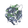
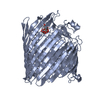
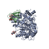
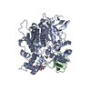
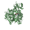
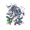
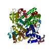
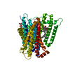
| Title | Crystal structure of ferric citrate transporter FecA in complex with iron-free citrate | ||||||
 Components Components | Iron(III) dicitrate transport protein fecA precursor | ||||||
 Keywords Keywords | MEMBRANE PROTEIN / outer membrane protein / beta barrel / TonB-dependent transport / citrate / siderophore | ||||||
| Function / homology |  Function and homology information Function and homology informationresponse to iron ion starvation / siderophore-iron transmembrane transporter activity / Metal ion assimilation from the host / siderophore-iron import into cell / siderophore transport / signal transduction involved in regulation of gene expression / transmembrane transporter complex / cell outer membrane / signaling receptor activity / intracellular iron ion homeostasis ...response to iron ion starvation / siderophore-iron transmembrane transporter activity / Metal ion assimilation from the host / siderophore-iron import into cell / siderophore transport / signal transduction involved in regulation of gene expression / transmembrane transporter complex / cell outer membrane / signaling receptor activity / intracellular iron ion homeostasis / regulation of DNA-templated transcription / membrane Similarity search - Function | ||||||
| Biological species |  | ||||||
| Method |  X-RAY DIFFRACTION / SYNCHROTRON / MOLECULAR REPLACEMENT / Resolution: 2.15 Å X-RAY DIFFRACTION / SYNCHROTRON / MOLECULAR REPLACEMENT / Resolution: 2.15 Å | ||||||
 Authors Authors | Yue, W.W. / Grizot, S. / Buchanan, S.K. | ||||||
 Citation Citation | Journal: J.Mol.Biol. / Year: 2003 Title: Structural evidence for iron-free citrate and ferric citrate binding to the TonB-dependent outer membrane transporter FecA Authors: Yue, W.W. / Grizot, S. / Buchanan, S.K. | ||||||
| History |
|
- Structure visualization
| Structure viewer | Molecule: MolmilJmol/JSmol |
|---|
- Downloads & links
-Download
| PDBx/mmCIF format | 1po0.cif.gz | 149 KB | Display | PDBx/mmCIF format |
|---|---|---|---|---|
| PDB format | pdb1po0.ent.gz | 113.3 KB | Display | PDB format |
| PDBx/mmJSON format | 1po0.json.gz | Tree view | PDBx/mmJSON format | |
| Others |  Other downloads Other downloads |
-Validation report
| Arichive directory | https://data.pdbj.org/pub/pdb/validation_reports/po/1po0ftp://data.pdbj.org/pub/pdb/validation_reports/po/1po0 | HTTPS FTP |
|---|
-Related structure data
| Related structure data | 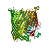 1pnzC  1po3C 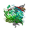 1kmoS C: citing same article ( S: Starting model for refinement |
|---|---|
| Similar structure data |


-Links
 PDBj
PDBj


- Assembly
| Deposited unit | 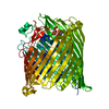
| ||||||||
|---|---|---|---|---|---|---|---|---|---|
| 1 |
| ||||||||
| Unit cell |
| ||||||||
| Details | The biological assembly is a monomer |
-Components
| #1: Protein | Mass: 83169.328 Da / Num. of mol.: 1 / Fragment: FecA residues 81-741 Source method: isolated from a genetically manipulated source Source: (gene. exp.) | ||
|---|---|---|---|
| #2: Chemical |   Mass: 189.100 Da / Num. of mol.: 2 / Fragment: iron-free dicitrate / Source method: obtained synthetically / Formula: C6H5O7 Mass: 189.100 Da / Num. of mol.: 2 / Fragment: iron-free dicitrate / Source method: obtained synthetically / Formula: C6H5O7#3: Water | ChemComp-HOH / |  Mass: 18.015 Da / Num. of mol.: 256 / Fragment: water / Source method: isolated from a natural source / Formula: H2O Mass: 18.015 Da / Num. of mol.: 256 / Fragment: water / Source method: isolated from a natural source / Formula: H2O |
-Experimental details
-Experiment
| Experiment | Method: X-RAY DIFFRACTION / Number of used crystals: 1 |
|---|
- Sample preparation
| Crystal | Density Matthews: 2.91 Å3/Da / Density % sol: 57.68 % | |||||||||||||||||||||||||||||||||||||||||||||||||||||||||||||||
|---|---|---|---|---|---|---|---|---|---|---|---|---|---|---|---|---|---|---|---|---|---|---|---|---|---|---|---|---|---|---|---|---|---|---|---|---|---|---|---|---|---|---|---|---|---|---|---|---|---|---|---|---|---|---|---|---|---|---|---|---|---|---|---|---|
| Crystal grow | Temperature: 295 K / Method: vapor diffusion, hanging drop / pH: 5.2 Details: PEG1000, LDAO, sodium citrate, sodium chloride , pH 5.2, VAPOR DIFFUSION, HANGING DROP, temperature 295K | |||||||||||||||||||||||||||||||||||||||||||||||||||||||||||||||
| Crystal grow | *PLUS Temperature: 22 ℃ / pH: 8 / Method: vapor diffusion, hanging drop | |||||||||||||||||||||||||||||||||||||||||||||||||||||||||||||||
| Components of the solutions | *PLUS
|
-Data collection
| Diffraction | Mean temperature: 100 K |
|---|---|
| Diffraction source | Source: SYNCHROTRON / Site: ESRF  / Beamline: ID14-1 / Wavelength: 0.918 Å / Beamline: ID14-1 / Wavelength: 0.918 Å |
| Detector | Type: ADSC QUANTUM 4 / Detector: CCD / Date: Feb 5, 2001 |
| Radiation | Monochromator: synchrotron / Protocol: SINGLE WAVELENGTH / Monochromatic (M) / Laue (L): M / Scattering type: x-ray |
| Radiation wavelength | Wavelength: 0.918 Å / Relative weight: 1 |
| Reflection | Resolution: 2.15→29.06 Å / Num. all: 54025 / Num. obs: 52397 / % possible obs: 98.1 % / Observed criterion σ(F): 1 / Observed criterion σ(I): 1 / Redundancy: 4 % / Biso Wilson estimate: 23.3 Å2 / Rmerge(I) obs: 0.112 / Net I/σ(I): 10.5 |
| Reflection shell | Resolution: 2.15→2.2 Å / Redundancy: 2.3 % / Rmerge(I) obs: 0.41 / Mean I/σ(I) obs: 2.3 / Num. unique all: 3498 / % possible all: 90.1 |
| Reflection | *PLUS Lowest resolution: 30 Å / Num. obs: 54094 / % possible obs: 98 % / Num. measured all: 215425 |
| Reflection shell | *PLUS % possible obs: 90.1 % / Num. unique obs: 3498 / Num. measured obs: 7930 / Rmerge(I) obs: 0.412 |
- Processing
| Software |
| ||||||||||||||||||||||||||||||||||||||||||||||||||||||||||||||||||||||||||||||||
|---|---|---|---|---|---|---|---|---|---|---|---|---|---|---|---|---|---|---|---|---|---|---|---|---|---|---|---|---|---|---|---|---|---|---|---|---|---|---|---|---|---|---|---|---|---|---|---|---|---|---|---|---|---|---|---|---|---|---|---|---|---|---|---|---|---|---|---|---|---|---|---|---|---|---|---|---|---|---|---|---|---|
| Refinement | Method to determine structure: MOLECULAR REPLACEMENT Starting model: PDB accession code 1KMO Resolution: 2.15→19.93 Å / Rfactor Rfree error: 0.005 / Isotropic thermal model: RESTRAINED / Cross valid method: THROUGHOUT / σ(F): 0 / Stereochemistry target values: Engh & Huber Details: Side chains were not built for residues ASP255, GLU293, GLN428, GLN695 and MET696
| ||||||||||||||||||||||||||||||||||||||||||||||||||||||||||||||||||||||||||||||||
| Solvent computation | Solvent model: FLAT MODEL / Bsol: 41.0399 Å2 / ksol: 0.342783 e/Å3 | ||||||||||||||||||||||||||||||||||||||||||||||||||||||||||||||||||||||||||||||||
| Displacement parameters | Biso mean: 34.8 Å2
| ||||||||||||||||||||||||||||||||||||||||||||||||||||||||||||||||||||||||||||||||
| Refine analyze |
| ||||||||||||||||||||||||||||||||||||||||||||||||||||||||||||||||||||||||||||||||
| Refinement step | Cycle: LAST / Resolution: 2.15→19.93 Å
| ||||||||||||||||||||||||||||||||||||||||||||||||||||||||||||||||||||||||||||||||
| Refine LS restraints |
| ||||||||||||||||||||||||||||||||||||||||||||||||||||||||||||||||||||||||||||||||
| LS refinement shell | Resolution: 2.15→2.28 Å / Rfactor Rfree error: 0.02 / Total num. of bins used: 6
| ||||||||||||||||||||||||||||||||||||||||||||||||||||||||||||||||||||||||||||||||
| Xplor file |
| ||||||||||||||||||||||||||||||||||||||||||||||||||||||||||||||||||||||||||||||||
| Refinement | *PLUS Lowest resolution: 20 Å / % reflection Rfree: 5 % | ||||||||||||||||||||||||||||||||||||||||||||||||||||||||||||||||||||||||||||||||
| Solvent computation | *PLUS | ||||||||||||||||||||||||||||||||||||||||||||||||||||||||||||||||||||||||||||||||
| Displacement parameters | *PLUS | ||||||||||||||||||||||||||||||||||||||||||||||||||||||||||||||||||||||||||||||||
| Refine LS restraints | *PLUS
|
