 Movie
Movie Controller


+ Open data

- Basic information
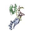
| Entry | Database: PDB / ID: 1pnn | |||||||||
|---|---|---|---|---|---|---|---|---|---|---|
| Title | PEPTIDE NUCLEIC ACID (PNA) COMPLEXED WITH DNA | |||||||||
 Components Components |
| |||||||||
 Keywords Keywords | PEPTIDE NUCLEIC ACID/DNA / HAIRPIN PNA:DNA TRIPLEX / TRIPLEX WATSON-CRICK HOOGSTEEN / PEPTIDE NUCLEIC ACID-DNA COMPLEX | |||||||||
| Function / homology | DNA / DNA (> 10) Function and homology information Function and homology information | |||||||||
| Method |  X-RAY DIFFRACTION / SYNCHROTRON / Resolution: 2.5 Å X-RAY DIFFRACTION / SYNCHROTRON / Resolution: 2.5 Å | |||||||||
 Authors Authors | Betts, L. / Veal, J.M. | |||||||||
 Citation Citation | Journal: Science / Year: 1995 Title: A Nucleic Acid Triple Helix Formed by a Peptide Nucleic Acid-DNA Complex Authors: Betts, L. / Josey, J.A. / Veal, J.M. / Jordan, S.R. #1: Journal: Science / Year: 1994Title: NMR Solution Structure of a Peptide Nucleic Acid Complexed with RNA Authors: Brown, S.C. / Thomson, S.A. / Veal, J.M. / Davis, D.G. | |||||||||
| History |
|
- Structure visualization
| Structure viewer | Molecule: MolmilJmol/JSmol |
|---|
- Downloads & links
-Download
| PDBx/mmCIF format | 1pnn.cif.gz | 58 KB | Display | PDBx/mmCIF format |
|---|---|---|---|---|
| PDB format | pdb1pnn.ent.gz | 49.5 KB | Display | PDB format |
| PDBx/mmJSON format | 1pnn.json.gz | Tree view | PDBx/mmJSON format | |
| Others |  Other downloads Other downloads |
-Validation report
| Arichive directory | https://data.pdbj.org/pub/pdb/validation_reports/pn/1pnnftp://data.pdbj.org/pub/pdb/validation_reports/pn/1pnn | HTTPS FTP |
|---|
-Related structure data
| Similar structure data |
|---|


-Links
 PDBj
PDBj


- Assembly
| Deposited unit | 
| ||||||||
|---|---|---|---|---|---|---|---|---|---|
| 1 |
| ||||||||
| Unit cell |
| ||||||||
| Noncrystallographic symmetry (NCS) | NCS oper: (Code: given Matrix: (-0.545117, -0.361267, -0.756527), Vector: |
-Components
| #1: DNA chain | Mass: 5322.071 Da / Num. of mol.: 2 / Source method: obtained synthetically #2: DNA chain | Mass: 2837.900 Da / Num. of mol.: 2 / Source method: obtained synthetically / Details: CRYSTALLIZED FROM AMMONIUM SULFATE AND TRIS PH 8.5 #3: Water | ChemComp-HOH / |  Mass: 18.015 Da / Num. of mol.: 35 / Source method: isolated from a natural source / Formula: H2O Mass: 18.015 Da / Num. of mol.: 35 / Source method: isolated from a natural source / Formula: H2O |
|---|
-Experimental details
-Experiment
| Experiment | Method: X-RAY DIFFRACTION |
|---|
- Sample preparation
| Crystal | Density Matthews: 3.31 Å3/Da / Density % sol: 62.79 % |
|---|---|
| Crystal grow | pH: 8.5 / Details: pH 8.5 |
-Data collection
| Diffraction |
| |||||||||||||||
|---|---|---|---|---|---|---|---|---|---|---|---|---|---|---|---|---|
| Diffraction source |
| |||||||||||||||
| Detector |
| |||||||||||||||
| Radiation |
| |||||||||||||||
| Radiation wavelength |
| |||||||||||||||
| Reflection | Resolution: 2.5→20 Å / Num. obs: 8374 / % possible obs: 93 % / Observed criterion σ(I): 1 / Redundancy: 3 % / Rmerge(I) obs: 0.043 |

- Processing
| Software |
| ||||||||||||||||||||||||||||||||||||||||||||||||||||||||||||
|---|---|---|---|---|---|---|---|---|---|---|---|---|---|---|---|---|---|---|---|---|---|---|---|---|---|---|---|---|---|---|---|---|---|---|---|---|---|---|---|---|---|---|---|---|---|---|---|---|---|---|---|---|---|---|---|---|---|---|---|---|---|
| Refinement | Resolution: 2.5→6 Å / σ(F): 2
| ||||||||||||||||||||||||||||||||||||||||||||||||||||||||||||
| Refine analyze | Luzzati coordinate error obs: 0.25 Å | ||||||||||||||||||||||||||||||||||||||||||||||||||||||||||||
| Refinement step | Cycle: LAST / Resolution: 2.5→6 Å
| ||||||||||||||||||||||||||||||||||||||||||||||||||||||||||||
| Refine LS restraints |
|
