 Movie
Movie Controller

[English] 日本語
 Yorodumi
- PDB-1pja: The crystal structure of palmitoyl protein thioesterase-2 reveals... -
+ Open data

- Basic information
| Entry | Database: PDB / ID: 1pja | ||||||
|---|---|---|---|---|---|---|---|
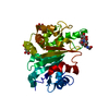
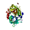
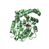
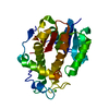
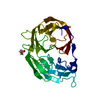
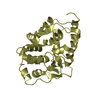
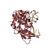
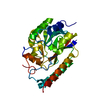
| Title | The crystal structure of palmitoyl protein thioesterase-2 reveals the basis for divergent substrate specificities of the two lysosomal thioesterases (PPT1 and PPT2) | ||||||
 Components Components | Palmitoyl-protein thioesterase 2 precursor | ||||||
 Keywords Keywords | HYDROLASE / glycoprotein / lysosome | ||||||
| Function / homology |  Function and homology information Function and homology informationpalmitoyl hydrolase activity / thiolester hydrolase activity / palmitoyl-CoA hydrolase / fatty-acyl-CoA biosynthetic process / fatty acyl-CoA hydrolase activity / Fatty acyl-CoA biosynthesis / lysosomal lumen / lysosome / extracellular exosome / extracellular region Similarity search - Function | ||||||
| Biological species |  Homo sapiens (human) Homo sapiens (human) | ||||||
| Method |  X-RAY DIFFRACTION / SYNCHROTRON / MOLECULAR REPLACEMENT / Resolution: 2.7 Å X-RAY DIFFRACTION / SYNCHROTRON / MOLECULAR REPLACEMENT / Resolution: 2.7 Å | ||||||
 Authors Authors | Calero, G. / Gupta, P. / Nonato, M.C. / Tandel, S. / Biehl, E.R. / Hofmann, S.L. / Clardy, J. | ||||||
 Citation Citation | Journal: J.Biol.Chem. / Year: 2003 Title: The crystal structure of palmitoyl protein thioesterase-2 (PPT2) reveals the basis for divergent substrate specificities of the two lysosomal thioesterases, PPT1 and PPT2. Authors: Calero, G. / Gupta, P. / Nonato, M.C. / Tandel, S. / Biehl, E.R. / Hofmann, S.L. / Clardy, J. | ||||||
| History |
|
- Structure visualization
| Structure viewer | Molecule: MolmilJmol/JSmol |
|---|
- Downloads & links
-Download
| PDBx/mmCIF format | 1pja.cif.gz | 68 KB | Display | PDBx/mmCIF format |
|---|---|---|---|---|
| PDB format | pdb1pja.ent.gz | 50.4 KB | Display | PDB format |
| PDBx/mmJSON format | 1pja.json.gz | Tree view | PDBx/mmJSON format | |
| Others |  Other downloads Other downloads |
-Validation report
| Arichive directory | https://data.pdbj.org/pub/pdb/validation_reports/pj/1pjaftp://data.pdbj.org/pub/pdb/validation_reports/pj/1pja | HTTPS FTP |
|---|
-Related structure data
| Related structure data | |
|---|---|
| Similar structure data |


-Links
 PDBj
PDBj- Assembly
| Deposited unit | 
| ||||||||
|---|---|---|---|---|---|---|---|---|---|
| 1 |
| ||||||||
| Unit cell |
|
-Components
| #1: Protein | Mass: 34342.289 Da / Num. of mol.: 1 Source method: isolated from a genetically manipulated source Source: (gene. exp.) Homo sapiens (human) / Gene: PPT2 / Production host:   Spodoptera frugiperda (fall armyworm) / References: UniProt: Q9UMR5, palmitoyl[protein] hydrolase Spodoptera frugiperda (fall armyworm) / References: UniProt: Q9UMR5, palmitoyl[protein] hydrolase | ||||
|---|---|---|---|---|---|
| #2: Sugar | ChemComp-NAG /   Type: D-saccharide, beta linking / Mass: 221.208 Da / Num. of mol.: 1 Type: D-saccharide, beta linking / Mass: 221.208 Da / Num. of mol.: 1Source method: isolated from a genetically manipulated source Formula: C8H15NO6 | ||||
| #3: Chemical |   Mass: 92.094 Da / Num. of mol.: 3 / Source method: obtained synthetically / Formula: C3H8O3 Mass: 92.094 Da / Num. of mol.: 3 / Source method: obtained synthetically / Formula: C3H8O3#4: Water | ChemComp-HOH / |  Mass: 18.015 Da / Num. of mol.: 50 / Source method: isolated from a natural source / Formula: H2O Mass: 18.015 Da / Num. of mol.: 50 / Source method: isolated from a natural source / Formula: H2OHas protein modification | Y | |
-Experimental details
-Experiment
| Experiment | Method: X-RAY DIFFRACTION / Number of used crystals: 1 |
|---|
- Sample preparation
| Crystal grow | Temperature: 277 K / Method: vapor diffusion, hanging drop / pH: 6 Details: 1.8-2.2M ammonium sulfate, 8% methyl-pentane-diol, 100 mM MES , pH 6.0, VAPOR DIFFUSION, HANGING DROP, temperature 277K | ||||||||||||||||||||||||||||||
|---|---|---|---|---|---|---|---|---|---|---|---|---|---|---|---|---|---|---|---|---|---|---|---|---|---|---|---|---|---|---|---|
| Crystal grow | *PLUS Temperature: 4 ℃ / Method: vapor diffusion, sitting drop / PH range low: 6.5 / PH range high: 5.5 | ||||||||||||||||||||||||||||||
| Components of the solutions | *PLUS
|
-Data collection
| Diffraction source | Source: SYNCHROTRON / Site: CHESS  / Beamline: A1 / Wavelength: 0.943 Å / Beamline: A1 / Wavelength: 0.943 Å |
|---|---|
| Detector | Type: ADSC QUANTUM 4 / Detector: CCD / Details: mirrors |
| Radiation | Protocol: SINGLE WAVELENGTH / Monochromatic (M) / Laue (L): M / Scattering type: x-ray |
| Radiation wavelength | Wavelength: 0.943 Å / Relative weight: 1 |
| Reflection | Resolution: 2.7→40 Å / Num. all: 34721 / Num. obs: 34721 / % possible obs: 99.8 % / Observed criterion σ(F): 2 / Observed criterion σ(I): 2 / Redundancy: 6.5 % / Rsym value: 0.046 / Net I/σ(I): 15.7 |
| Reflection shell | Highest resolution: 2.7 Å / Mean I/σ(I) obs: 2.5 |
| Reflection | *PLUS % possible obs: 100 % / Rmerge(I) obs: 0.046 |
- Processing
| Software |
| ||||||||||||||||||
|---|---|---|---|---|---|---|---|---|---|---|---|---|---|---|---|---|---|---|---|
| Refinement | Method to determine structure: MOLECULAR REPLACEMENT / Resolution: 2.7→40 Å / σ(F): 0
| ||||||||||||||||||
| Refinement step | Cycle: LAST / Resolution: 2.7→40 Å
| ||||||||||||||||||
| Refinement | *PLUS % reflection Rfree: 5 % / Rfactor Rfree: 0.245 / Rfactor Rwork: 0.225 | ||||||||||||||||||
| Solvent computation | *PLUS | ||||||||||||||||||
| Displacement parameters | *PLUS | ||||||||||||||||||
| Refine LS restraints | *PLUS
|
