 Movie
Movie Controller


+ Open data

- Basic information
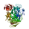
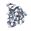
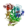
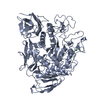
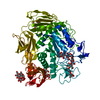
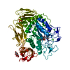
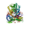
| Entry | Database: PDB / ID: 1pj9 | |||||||||
|---|---|---|---|---|---|---|---|---|---|---|
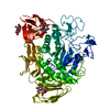
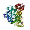
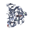
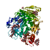
| Title | Bacillus circulans strain 251 loop mutant 183-195 | |||||||||
 Components Components | Cyclomaltodextrin glucanotransferase | |||||||||
 Keywords Keywords | TRANSFERASE / glycosyltransferase / cyclodextrin | |||||||||
| Function / homology |  Function and homology information Function and homology informationcyclomaltodextrin glucanotransferase / cyclomaltodextrin glucanotransferase activity / starch binding / alpha-amylase activity / carbohydrate metabolic process / extracellular region / metal ion binding Similarity search - Function | |||||||||
| Biological species |  Bacillus circulans (bacteria) Bacillus circulans (bacteria) | |||||||||
| Method |  X-RAY DIFFRACTION / FOURIER SYNTHESIS / Resolution: 2 Å X-RAY DIFFRACTION / FOURIER SYNTHESIS / Resolution: 2 Å | |||||||||
 Authors Authors | Rozeboom, H.J. / Dijkstra, B.W. | |||||||||
 Citation Citation | Journal: PROTEINS: STRUCT.,FUNCT.,GENET. / Year: 2004 Title: Improved thermostability of bacillus circulans cyclodextrin glycosyltransferase by the introduction of a salt bridge Authors: Leemhuis, H. / Rozeboom, H.J. / Dijkstra, B.W. / Dijkhuizen, L. | |||||||||
| History |
|
- Structure visualization
| Structure viewer | Molecule: MolmilJmol/JSmol |
|---|
- Downloads & links
-Download
| PDBx/mmCIF format | 1pj9.cif.gz | 168.8 KB | Display | PDBx/mmCIF format |
|---|---|---|---|---|
| PDB format | pdb1pj9.ent.gz | 129.9 KB | Display | PDB format |
| PDBx/mmJSON format | 1pj9.json.gz | Tree view | PDBx/mmJSON format | |
| Others |  Other downloads Other downloads |
-Validation report
| Arichive directory | https://data.pdbj.org/pub/pdb/validation_reports/pj/1pj9ftp://data.pdbj.org/pub/pdb/validation_reports/pj/1pj9 | HTTPS FTP |
|---|
-Related structure data
| Related structure data | 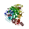 1d3cS S: Starting model for refinement |
|---|---|
| Similar structure data |
-Links
 PDBj
PDBj


- Assembly
| Deposited unit | 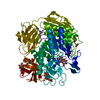
| ||||||||
|---|---|---|---|---|---|---|---|---|---|
| 1 |
| ||||||||
| Unit cell |
|
-Components
-Protein , 1 types, 1 molecules A
| #1: Protein | Mass: 74649.461 Da / Num. of mol.: 1 / Mutation: T185S, T186Y, N188D, K192R Source method: isolated from a genetically manipulated source Source: (gene. exp.) Bacillus circulans (bacteria) / Strain: 251 / Plasmid: pDP66k- / Production host: References: UniProt: P43379, cyclomaltodextrin glucanotransferase |
|---|
-Sugars , 3 types, 6 molecules 
| #2: Polysaccharide |   Source method: isolated from a genetically manipulated source Details: oligosaccharide / References: alpha-maltose #3: Polysaccharide |   Source method: isolated from a genetically manipulated source Details: oligosaccharide / References: alpha-maltotriose #4: Sugar | ChemComp-GLC / |  Type: D-saccharide, alpha linking / Mass: 180.156 Da / Num. of mol.: 1 Type: D-saccharide, alpha linking / Mass: 180.156 Da / Num. of mol.: 1Source method: isolated from a genetically manipulated source Formula: C6H12O6 |
|---|
-Non-polymers , 4 types, 805 molecules 


| #5: Chemical | ChemComp-MPD / ( Mass: 118.174 Da / Num. of mol.: 1 / Source method: obtained synthetically / Formula: C6H14O2 / Comment: precipitant*YM Mass: 118.174 Da / Num. of mol.: 1 / Source method: obtained synthetically / Formula: C6H14O2 / Comment: precipitant*YM | ||||
|---|---|---|---|---|---|
| #6: Chemical |  Mass: 60.052 Da / Num. of mol.: 3 / Source method: obtained synthetically / Formula: C2H4O2 Mass: 60.052 Da / Num. of mol.: 3 / Source method: obtained synthetically / Formula: C2H4O2#7: Chemical |  Mass: 40.078 Da / Num. of mol.: 3 / Source method: obtained synthetically / Formula: Ca Mass: 40.078 Da / Num. of mol.: 3 / Source method: obtained synthetically / Formula: Ca#8: Water | ChemComp-HOH / | Mass: 18.015 Da / Num. of mol.: 798 / Source method: isolated from a natural source / Formula: H2O |
-Details
| Has protein modification | Y |
|---|
-Experimental details
-Experiment
| Experiment | Method: X-RAY DIFFRACTION / Number of used crystals: 1 |
|---|
- Sample preparation
| Crystal | Density Matthews: 2.81 Å3/Da / Density % sol: 56.27 % | ||||||||||||||||||||||||
|---|---|---|---|---|---|---|---|---|---|---|---|---|---|---|---|---|---|---|---|---|---|---|---|---|---|
| Crystal grow | Temperature: 295 K / Method: vapor diffusion, hanging drop / pH: 7.5 Details: MPD, Ca, HEPES, maltose, pH 7.5, VAPOR DIFFUSION, HANGING DROP, temperature 295K | ||||||||||||||||||||||||
| Crystal grow | *PLUS Method: vapor diffusion / Details: Lawson, C.L., (1994) J. Mol. Biol., 236, 590. | ||||||||||||||||||||||||
| Components of the solutions | *PLUS
|
-Data collection
| Diffraction | Mean temperature: 120 K |
|---|---|
| Diffraction source | Source: ROTATING ANODE / Type: ENRAF-NONIUS / Wavelength: 1.5418 Å |
| Detector | Type: MARRESEARCH / Detector: CCD / Date: Aug 23, 2002 / Details: Osmic mirrors |
| Radiation | Protocol: SINGLE WAVELENGTH / Monochromatic (M) / Laue (L): M / Scattering type: x-ray |
| Radiation wavelength | Wavelength: 1.5418 Å / Relative weight: 1 |
| Reflection | Resolution: 2→50 Å / Num. all: 57530 / Num. obs: 57530 / % possible obs: 98.3 % / Observed criterion σ(F): 0 / Observed criterion σ(I): 0 / Biso Wilson estimate: 12.1 Å2 / Rmerge(I) obs: 0.032 / Net I/σ(I): 34.7 |
| Reflection shell | Resolution: 2→2.07 Å / Rmerge(I) obs: 0.115 / Mean I/σ(I) obs: 7.8 / Num. unique all: 5528 / % possible all: 96.3 |
| Reflection | *PLUS Lowest resolution: 32 Å / Num. obs: 57077 / % possible obs: 98.8 % / Num. measured all: 363420 |
| Reflection shell | *PLUS % possible obs: 96.3 % / Mean I/σ(I) obs: 8.9 |
- Processing
| Software |
| ||||||||||||||||||||||||||||||||||||
|---|---|---|---|---|---|---|---|---|---|---|---|---|---|---|---|---|---|---|---|---|---|---|---|---|---|---|---|---|---|---|---|---|---|---|---|---|---|
| Refinement | Method to determine structure: FOURIER SYNTHESIS Starting model: PDB ENTRY 1D3C Resolution: 2→31.9 Å / Rfactor Rfree error: 0.003 / Data cutoff high absF: 2283082.17 / Data cutoff high rms absF: 2283082.17 / Data cutoff low absF: 0 / Isotropic thermal model: RESTRAINED / Cross valid method: THROUGHOUT / σ(F): 0 / Stereochemistry target values: Engh & Huber
| ||||||||||||||||||||||||||||||||||||
| Solvent computation | Solvent model: FLAT MODEL / Bsol: 76.6193 Å2 / ksol: 0.428511 e/Å3 | ||||||||||||||||||||||||||||||||||||
| Displacement parameters | Biso mean: 18.3 Å2
| ||||||||||||||||||||||||||||||||||||
| Refine analyze |
| ||||||||||||||||||||||||||||||||||||
| Refinement step | Cycle: LAST / Resolution: 2→31.9 Å
| ||||||||||||||||||||||||||||||||||||
| Refine LS restraints |
| ||||||||||||||||||||||||||||||||||||
| LS refinement shell | Resolution: 2→2.07 Å / Rfactor Rfree error: 0.014 / Total num. of bins used: 10
| ||||||||||||||||||||||||||||||||||||
| Xplor file |
| ||||||||||||||||||||||||||||||||||||
| Refinement | *PLUS Highest resolution: 2 Å / Lowest resolution: 32 Å | ||||||||||||||||||||||||||||||||||||
| Solvent computation | *PLUS | ||||||||||||||||||||||||||||||||||||
| Displacement parameters | *PLUS | ||||||||||||||||||||||||||||||||||||
| Refine LS restraints | *PLUS
| ||||||||||||||||||||||||||||||||||||
| LS refinement shell | *PLUS Rfactor Rfree: 0.224 |
