 Movie
Movie Controller

[English] 日本語
 Yorodumi
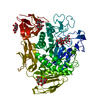
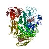
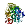
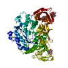
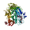
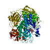
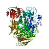
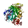
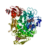
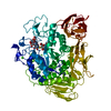
- PDB-1cxi: WILD-TYPE CGTASE FROM BACILLUS CIRCULANS STRAIN 251 AT 120 K AND ... -
+ Open data

- Basic information
| Entry | Database: PDB / ID: 1cxi | |||||||||
|---|---|---|---|---|---|---|---|---|---|---|
| Title | WILD-TYPE CGTASE FROM BACILLUS CIRCULANS STRAIN 251 AT 120 K AND PH 7.55 | |||||||||
 Components Components | CYCLODEXTRIN GLYCOSYLTRANSFERASE | |||||||||
 Keywords Keywords | GLYCOSYLTRANSFERASE | |||||||||
| Function / homology |  Function and homology information Function and homology informationcyclomaltodextrin glucanotransferase / cyclomaltodextrin glucanotransferase activity / starch binding / alpha-amylase activity / carbohydrate metabolic process / extracellular region / metal ion binding Similarity search - Function | |||||||||
| Biological species |  Bacillus circulans (bacteria) Bacillus circulans (bacteria) | |||||||||
| Method |  X-RAY DIFFRACTION / Resolution: 2.2 Å X-RAY DIFFRACTION / Resolution: 2.2 Å | |||||||||
 Authors Authors | Knegtel, R.M.A. / Strokopytov, B.V. / Dijkstra, B.W. | |||||||||
 Citation Citation | Journal: J.Biol.Chem. / Year: 1995 Title: Crystallographic studies of the interaction of cyclodextrin glycosyltransferase from Bacillus circulans strain 251 with natural substrates and products. Authors: Knegtel, R.M. / Strokopytov, B. / Penninga, D. / Faber, O.G. / Rozeboom, H.J. / Kalk, K.H. / Dijkhuizen, L. / Dijkstra, B.W. #1: Journal: Biochemistry / Year: 1995Title: X-Ray Structure of Cyclodextrin Glycosyltransferase Complexed with Acarbose. Implications for the Catalytic Mechanism of Glycosidases Authors: Strokopytov, B. / Penninga, D. / Rozeboom, H.J. / Kalk, K.H. / Dijkhuizen, L. / Dijkstra, B.W. #2: Journal: J.Mol.Biol. / Year: 1994Title: Nucleotide Sequence and X-Ray Structure of Cyclodextrin Glycosyltransferase from Bacillus Circulans Strain 251 in a Maltose-Dependent Crystal Form Authors: Lawson, C.L.L. / Van Montfort, R. / Strokopytov, B. / Rozeboom, H.J. / Kalk, K.H. / De Vries, G.E. / Penninga, D. / Dijkhuizen, L. / Dijkstra, B.W. #3: Journal: J.Mol.Biol. / Year: 1990Title: Maltodextrin-Dependent Crystallization of Cyclomalto-Dextrin Glucanotransferase from Bacillus Circulans Authors: Lawson, C.L.L. / Bergsma, J. / Bruinenberg, P.M. / De Vries, G. / Dijkhuizen, L. / Dijkstra, B.W. | |||||||||
| History |
|
- Structure visualization
| Structure viewer | Molecule: MolmilJmol/JSmol |
|---|
- Downloads & links
-Download
| PDBx/mmCIF format | 1cxi.cif.gz | 156.1 KB | Display | PDBx/mmCIF format |
|---|---|---|---|---|
| PDB format | pdb1cxi.ent.gz | 120.5 KB | Display | PDB format |
| PDBx/mmJSON format | 1cxi.json.gz | Tree view | PDBx/mmJSON format | |
| Others |  Other downloads Other downloads |
-Validation report
| Arichive directory | https://data.pdbj.org/pub/pdb/validation_reports/cx/1cxiftp://data.pdbj.org/pub/pdb/validation_reports/cx/1cxi | HTTPS FTP |
|---|
-Related structure data
-Links
 PDBj
PDBj


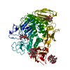
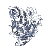
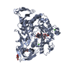
- Assembly
| Deposited unit | 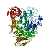
| ||||||||
|---|---|---|---|---|---|---|---|---|---|
| 1 |
| ||||||||
| Unit cell |
| ||||||||
| Atom site foot note | 1: CIS PROLINE - PRO 372 / 2: CIS PROLINE - PRO 506 / 3: CIS PROLINE - PRO 624 |
-Components
| #1: Protein | Mass: 74575.484 Da / Num. of mol.: 1 / Source method: isolated from a natural source / Source: (natural) Bacillus circulans (bacteria) / Strain: 251References: UniProt: P43379, cyclomaltodextrin glucanotransferase | ||||||
|---|---|---|---|---|---|---|---|
| #2: Polysaccharide |   Source method: isolated from a genetically manipulated source Details: oligosaccharide / References: alpha-maltose #3: Chemical |   Mass: 40.078 Da / Num. of mol.: 2 / Source method: obtained synthetically / Formula: Ca Mass: 40.078 Da / Num. of mol.: 2 / Source method: obtained synthetically / Formula: Ca#4: Water | ChemComp-HOH / |  Mass: 18.015 Da / Num. of mol.: 468 / Source method: isolated from a natural source / Formula: H2O Mass: 18.015 Da / Num. of mol.: 468 / Source method: isolated from a natural source / Formula: H2OHas protein modification | Y | |
-Experimental details
-Experiment
| Experiment | Method: X-RAY DIFFRACTION |
|---|
- Sample preparation
| Crystal | Density Matthews: 2.85 Å3/Da / Density % sol: 56.83 % | ||||||||||||||||||||||||||||||
|---|---|---|---|---|---|---|---|---|---|---|---|---|---|---|---|---|---|---|---|---|---|---|---|---|---|---|---|---|---|---|---|
| Crystal grow | pH: 7.55 / Details: pH 7.55 | ||||||||||||||||||||||||||||||
| Crystal grow | *PLUS pH: 5.5 / Method: vapor diffusion | ||||||||||||||||||||||||||||||
| Components of the solutions | *PLUS
|
-Data collection
| Diffraction | Mean temperature: 120 K |
|---|---|
| Diffraction source | Wavelength: 1.5418 |
| Detector | Type: ENRAF-NONIUS FAST / Detector: DIFFRACTOMETER / Date: Jul 2, 1992 |
| Radiation | Monochromatic (M) / Laue (L): M / Scattering type: x-ray |
| Radiation wavelength | Wavelength: 1.5418 Å / Relative weight: 1 |
| Reflection | Redundancy: 4.7 % / Rmerge(I) obs: 0.087 |
| Reflection | *PLUS Highest resolution: 2.2 Å / Lowest resolution: 9.6 Å / Num. obs: 39669 / % possible obs: 89.7 % / Num. measured all: 186267 / Rmerge(I) obs: 0.087 |
| Reflection shell | *PLUS Highest resolution: 2.2 Å / Lowest resolution: 2.23 Å / % possible obs: 79.3 % |
- Processing
| Software |
| ||||||||||||||||||||||||||||||
|---|---|---|---|---|---|---|---|---|---|---|---|---|---|---|---|---|---|---|---|---|---|---|---|---|---|---|---|---|---|---|---|
| Refinement | Resolution: 2.2→8 Å / σ(F): 0 /
| ||||||||||||||||||||||||||||||
| Refinement step | Cycle: LAST / Resolution: 2.2→8 Å
| ||||||||||||||||||||||||||||||
| Refine LS restraints |
| ||||||||||||||||||||||||||||||
| Software | *PLUS Name: TNT / Classification: refinement | ||||||||||||||||||||||||||||||
| Refinement | *PLUS % reflection Rfree: 10 % / Rfactor Rfree: 0.243 | ||||||||||||||||||||||||||||||
| Solvent computation | *PLUS | ||||||||||||||||||||||||||||||
| Displacement parameters | *PLUS Biso mean: 12.1 Å2 | ||||||||||||||||||||||||||||||
| Refine LS restraints | *PLUS
|
