 Movie
Movie Controller


+ Open data

- Basic information
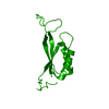
| Entry | Database: PDB / ID: 1pil | ||||||
|---|---|---|---|---|---|---|---|
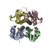
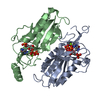
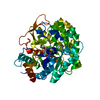
| Title | STRUCTURE OF THE ESCHERICHIA COLI SIGNAL TRANSDUCING PROTEIN PII | ||||||
 Components Components | SIGNAL TRANSDUCING PROTEIN P2 | ||||||
 Keywords Keywords | NITROGEN REGULATORY PROTEIN | ||||||
| Function / homology |  Function and homology information Function and homology informationregulation of nitrogen utilization / regulation of fatty acid biosynthetic process / small molecule binding / enzyme regulator activity / enzyme activator activity / DNA-templated transcription / ATP binding / identical protein binding / cytosol Similarity search - Function | ||||||
| Biological species |  | ||||||
| Method |  X-RAY DIFFRACTION / Resolution: 2.7 Å X-RAY DIFFRACTION / Resolution: 2.7 Å | ||||||
 Authors Authors | Ollis, D.L. / Cheah, U.E. / Carr, P.D. / Suffolk, P.M. | ||||||
 Citation Citation | Journal: Structure / Year: 1994 Title: Structure of the Escherichia coli signal transducing protein PII. Authors: Cheah, E. / Carr, P.D. / Suffolk, P.M. / Vasudevan, S.G. / Dixon, N.E. / Ollis, D.L. #1: Journal: FEBS Lett. / Year: 1994Title: Escherichia Coli Pii Protein: Purification, Crystallization, and Oligomeric Structure Authors: Vasudevan, S.G. / Gedye, C. / Dixon, N.E. / Cheah, U.E. / Carr, P.D. / Suffolk, P.M. / Jeffrey, P.D. / Ollis, D.L. | ||||||
| History |
|
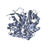
- Structure visualization
| Structure viewer | Molecule: MolmilJmol/JSmol |
|---|
- Downloads & links
-Download
| PDBx/mmCIF format | 1pil.cif.gz | 28.8 KB | Display | PDBx/mmCIF format |
|---|---|---|---|---|
| PDB format | pdb1pil.ent.gz | 19.6 KB | Display | PDB format |
| PDBx/mmJSON format | 1pil.json.gz | Tree view | PDBx/mmJSON format | |
| Others |  Other downloads Other downloads |
-Validation report
| Arichive directory | https://data.pdbj.org/pub/pdb/validation_reports/pi/1pilftp://data.pdbj.org/pub/pdb/validation_reports/pi/1pil | HTTPS FTP |
|---|
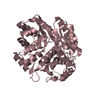
-Related structure data
| Similar structure data |
|---|


-Links
 PDBj
PDBj- Assembly
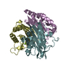
| Deposited unit | 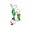
| ||||||||
|---|---|---|---|---|---|---|---|---|---|
| 1 | 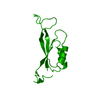
| ||||||||
| Unit cell |
| ||||||||
| Details | THE TWO TRANSFORMATIONS PRESENTED BELOW WILL GENERATE A TRIMER FROM THE SUBMITTED COORDINATES. THEY ARE CRYSTALLOGRAPHIC TRANSFORMATIONS. SYMTY1 1 -0.500000 0.866030 0.000000 -30.81050 SYMTY2 1 -0.866030 -0.500000 0.000000 53.36540 SYMTY3 1 0.000000 0.000000 1.000000 0.00000 SYMTY1 2 -0.500000 -0.866030 0.000000 30.81050 SYMTY2 2 0.866030 -0.500000 0.000000 53.36540 SYMTY3 2 0.000000 0.000000 1.000000 0.00000 |
-Components
| #1: Protein | Mass: 12443.442 Da / Num. of mol.: 1 Source method: isolated from a genetically manipulated source Source: (gene. exp.) |
|---|
-Experimental details
-Experiment
| Experiment | Method: X-RAY DIFFRACTION |
|---|
- Sample preparation
| Crystal | Density Matthews: 2.48 Å3/Da / Density % sol: 50.35 % | ||||||||||||||||||||
|---|---|---|---|---|---|---|---|---|---|---|---|---|---|---|---|---|---|---|---|---|---|
| Crystal grow | *PLUS Temperature: 4 ℃ / pH: 7 / Method: vapor diffusion, hanging drop / Details: Vasudevan, S.G., (1994) FEBS Lett., 337, 255. | ||||||||||||||||||||
| Components of the solutions | *PLUS
|
-Data collection
| Radiation | Scattering type: x-ray |
|---|---|
| Radiation wavelength | Relative weight: 1 |
| Reflection | *PLUS Highest resolution: 2.2 Å / Num. obs: 6578 / % possible obs: 89.5 % / Rmerge(I) obs: 0.025 |
- Processing
| Software |
| ||||||||||||||||||||||||||||||||||||||||||||||||||||||||||||
|---|---|---|---|---|---|---|---|---|---|---|---|---|---|---|---|---|---|---|---|---|---|---|---|---|---|---|---|---|---|---|---|---|---|---|---|---|---|---|---|---|---|---|---|---|---|---|---|---|---|---|---|---|---|---|---|---|---|---|---|---|---|
| Refinement | Rfactor Rwork: 0.24 / Rfactor obs: 0.24 / Highest resolution: 2.7 Å | ||||||||||||||||||||||||||||||||||||||||||||||||||||||||||||
| Refinement step | Cycle: LAST / Highest resolution: 2.7 Å
| ||||||||||||||||||||||||||||||||||||||||||||||||||||||||||||
| Refine LS restraints |
| ||||||||||||||||||||||||||||||||||||||||||||||||||||||||||||
| Refinement | *PLUS Lowest resolution: 10 Å / Rfactor obs: 0.245 / Rfactor Rwork: 0.245 | ||||||||||||||||||||||||||||||||||||||||||||||||||||||||||||
| Solvent computation | *PLUS | ||||||||||||||||||||||||||||||||||||||||||||||||||||||||||||
| Displacement parameters | *PLUS |
