 Movie
Movie Controller

[English] 日本語
 Yorodumi
- PDB-1pi6: YEAST ACTIN INTERACTING PROTEIN 1 (Aip1), ORTHORHOMBIC CRYSTAL FORM -
+ Open data

- Basic information
| Entry | Database: PDB / ID: 1pi6 | ||||||
|---|---|---|---|---|---|---|---|
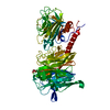
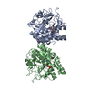
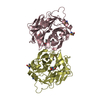
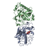
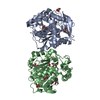
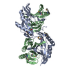
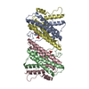
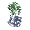
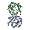
| Title | YEAST ACTIN INTERACTING PROTEIN 1 (Aip1), ORTHORHOMBIC CRYSTAL FORM | ||||||
 Components Components | Actin interacting protein 1 | ||||||
 Keywords Keywords | PROTEIN BINDING / WD repeat / beta-propeller | ||||||
| Function / homology |  Function and homology information Function and homology informationnegative regulation of cytokinesis / Platelet degranulation / actin cortical patch / barbed-end actin filament capping / actin filament depolymerization / mating projection tip / cortical actin cytoskeleton / actin filament / actin filament binding / actin binding ...negative regulation of cytokinesis / Platelet degranulation / actin cortical patch / barbed-end actin filament capping / actin filament depolymerization / mating projection tip / cortical actin cytoskeleton / actin filament / actin filament binding / actin binding / nucleus / cytoplasm Similarity search - Function | ||||||
| Biological species |  | ||||||
| Method |  X-RAY DIFFRACTION / SYNCHROTRON / MOLECULAR REPLACEMENT / Resolution: 2.5 Å X-RAY DIFFRACTION / SYNCHROTRON / MOLECULAR REPLACEMENT / Resolution: 2.5 Å | ||||||
 Authors Authors | Voegtli, W.C. / Madrona, A.Y. / Wilson, D.K. | ||||||
 Citation Citation | Journal: J.Biol.Chem. / Year: 2003 Title: The structure of Aip1p, a WD repeat protein that regulates Cofilin-mediated actin depolymerization. Authors: Voegtli, W.C. / Madrona, A.Y. / Wilson, D.K. | ||||||
| History |
|
- Structure visualization
| Structure viewer | Molecule: MolmilJmol/JSmol |
|---|
- Downloads & links
-Download
| PDBx/mmCIF format | 1pi6.cif.gz | 130.7 KB | Display | PDBx/mmCIF format |
|---|---|---|---|---|
| PDB format | pdb1pi6.ent.gz | 101.3 KB | Display | PDB format |
| PDBx/mmJSON format | 1pi6.json.gz | Tree view | PDBx/mmJSON format | |
| Others |  Other downloads Other downloads |
-Validation report
| Summary document | 1pi6_validation.pdf.gz | 369.2 KB | Display | wwPDB validaton report |
|---|---|---|---|---|
| Full document | 1pi6_full_validation.pdf.gz | 390.1 KB | Display | |
| Data in XML | 1pi6_validation.xml.gz | 15.6 KB | Display | |
| Data in CIF | 1pi6_validation.cif.gz | 23.6 KB | Display | |
| Arichive directory | https://data.pdbj.org/pub/pdb/validation_reports/pi/1pi6ftp://data.pdbj.org/pub/pdb/validation_reports/pi/1pi6 | HTTPS FTP |
-Related structure data
| Related structure data | 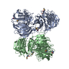 1pguSC S: Starting model for refinement C: citing same article ( |
|---|---|
| Similar structure data |

-Links
 PDBj
PDBj
- Assembly
| Deposited unit | 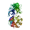
| ||||||||
|---|---|---|---|---|---|---|---|---|---|
| 1 |
| ||||||||
| 2 | 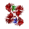
| ||||||||
| Unit cell |
|
-Components
| #1: Protein | Mass: 67415.289 Da / Num. of mol.: 1 / Mutation: H530R Source method: isolated from a genetically manipulated source Source: (gene. exp.) Gene: AIP1 OR YMR092C OR YM9582.17C / Plasmid: pTyb4 / Production host:  |
|---|---|
| #2: Chemical | ChemComp-ZN /   Mass: 65.409 Da / Num. of mol.: 1 / Source method: obtained synthetically / Formula: Zn Mass: 65.409 Da / Num. of mol.: 1 / Source method: obtained synthetically / Formula: Zn |
| #3: Water | ChemComp-HOH /  Mass: 18.015 Da / Num. of mol.: 132 / Source method: isolated from a natural source / Formula: H2O Mass: 18.015 Da / Num. of mol.: 132 / Source method: isolated from a natural source / Formula: H2O |
| Has protein modification | Y |
-Experimental details
-Experiment
| Experiment | Method: X-RAY DIFFRACTION / Number of used crystals: 1 |
|---|
- Sample preparation
| Crystal | Density Matthews: 2.46 Å3/Da / Density % sol: 49.97 % | |||||||||||||||||||||||||||||||||||
|---|---|---|---|---|---|---|---|---|---|---|---|---|---|---|---|---|---|---|---|---|---|---|---|---|---|---|---|---|---|---|---|---|---|---|---|---|
| Crystal grow | Temperature: 293 K / Method: vapor diffusion, hanging drop / pH: 7.5 Details: 15% PEG 4000, 100mM NaCl, 100mM HEPES, pH 7.5, VAPOR DIFFUSION, HANGING DROP, temperature 293K | |||||||||||||||||||||||||||||||||||
| Crystal grow | *PLUS Method: vapor diffusion, hanging drop | |||||||||||||||||||||||||||||||||||
| Components of the solutions | *PLUS
|
-Data collection
| Diffraction | Mean temperature: 100 K |
|---|---|
| Diffraction source | Source: SYNCHROTRON / Site: SSRL  / Beamline: BL9-1 / Wavelength: 0.97 Å / Beamline: BL9-1 / Wavelength: 0.97 Å |
| Detector | Type: MARRESEARCH / Detector: IMAGE PLATE / Date: Apr 20, 2001 |
| Radiation | Protocol: SINGLE WAVELENGTH / Monochromatic (M) / Laue (L): M / Scattering type: x-ray |
| Radiation wavelength | Wavelength: 0.97 Å / Relative weight: 1 |
| Reflection | Resolution: 2.5→25 Å / Num. all: 23703 / Num. obs: 21676 / % possible obs: 91.4 % / Observed criterion σ(F): 1 / Observed criterion σ(I): 1 / Biso Wilson estimate: 22.9 Å2 |
| Reflection shell | Resolution: 2.5→2.66 Å / % possible all: 80.6 |
| Reflection | *PLUS Highest resolution: 2.5 Å / Lowest resolution: 25 Å / Num. obs: 22190 / % possible obs: 93.5 % / Num. measured all: 192575 / Rmerge(I) obs: 0.063 |
| Reflection shell | *PLUS Lowest resolution: 2.54 Å / % possible obs: 83.5 % / Rmerge(I) obs: 0.235 |
- Processing
| Software |
| ||||||||||||||||||||||||||||||||||||
|---|---|---|---|---|---|---|---|---|---|---|---|---|---|---|---|---|---|---|---|---|---|---|---|---|---|---|---|---|---|---|---|---|---|---|---|---|---|
| Refinement | Method to determine structure: MOLECULAR REPLACEMENT Starting model: PDB ENTRY 1PGU Resolution: 2.5→24.82 Å / Rfactor Rfree error: 0.008 / Data cutoff high absF: 434259.93 / Data cutoff high rms absF: 434259.93 / Data cutoff low absF: 0 / Isotropic thermal model: RESTRAINED / Cross valid method: THROUGHOUT / σ(F): 0
| ||||||||||||||||||||||||||||||||||||
| Solvent computation | Solvent model: FLAT MODEL / Bsol: 14.5035 Å2 / ksol: 0.3143 e/Å3 | ||||||||||||||||||||||||||||||||||||
| Displacement parameters | Biso mean: 23.4 Å2
| ||||||||||||||||||||||||||||||||||||
| Refine analyze |
| ||||||||||||||||||||||||||||||||||||
| Refinement step | Cycle: LAST / Resolution: 2.5→24.82 Å
| ||||||||||||||||||||||||||||||||||||
| Refine LS restraints |
| ||||||||||||||||||||||||||||||||||||
| LS refinement shell | Resolution: 2.5→2.66 Å / Rfactor Rfree error: 0.025 / Total num. of bins used: 6
| ||||||||||||||||||||||||||||||||||||
| Xplor file |
| ||||||||||||||||||||||||||||||||||||
| Refinement | *PLUS Highest resolution: 2.5 Å / Lowest resolution: 25 Å / Rfactor Rfree: 0.263 / Rfactor Rwork: 0.22 | ||||||||||||||||||||||||||||||||||||
| Solvent computation | *PLUS | ||||||||||||||||||||||||||||||||||||
| Displacement parameters | *PLUS | ||||||||||||||||||||||||||||||||||||
| Refine LS restraints | *PLUS
|
