 Movie
Movie Controller

[English] 日本語
 Yorodumi
- PDB-1pcl: UNUSUAL STRUCTURAL FEATURES IN THE PARALLEL BETA-HELIX IN PECTATE... -
+ Open data

- Basic information
| Entry | Database: PDB / ID: 1pcl | ||||||
|---|---|---|---|---|---|---|---|
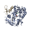
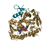
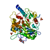
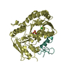
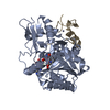
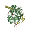
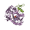
| Title | UNUSUAL STRUCTURAL FEATURES IN THE PARALLEL BETA-HELIX IN PECTATE LYASES | ||||||
 Components Components | PECTATE LYASE E | ||||||
 Keywords Keywords | LYASE (ACTING ON POLYSACCHARIDES) | ||||||
| Function / homology |  Function and homology information Function and homology informationpectate lyase / pectate lyase activity / pectin catabolic process / extracellular region / metal ion binding Similarity search - Function | ||||||
| Biological species |  Erwinia chrysanthemi (bacteria) Erwinia chrysanthemi (bacteria) | ||||||
| Method |  X-RAY DIFFRACTION / Resolution: 2.2 Å X-RAY DIFFRACTION / Resolution: 2.2 Å | ||||||
 Authors Authors | Lietzke, S.E. / Keen, N.T. / Jurnak, F. | ||||||
 Citation Citation | Journal: Structure / Year: 1993 Title: Unusual structural features in the parallel beta-helix in pectate lyases. Authors: Yoder, M.D. / Lietzke, S.E. / Jurnak, F. #1: Journal: Plant Physiol. / Year: 1996Title: The Refined Three-Dimensional Structure of Pectate Lyase E from Erwinia Chrysanthemi at 2.2 A Resolution. Authors: Lietzke, S.E. / Keen, N.T. / Jurnak, F. #2: Journal: J.Mol.Biol. / Year: 1989Title: Preliminary Crystallographic Analysis of a Plant Pathogenic Factor: Pectate Lyase Authors: Kim, C.-Y. / Mosser, V. / Keen, N. / Jurnak, F. #3: Journal: J.Bacteriol. / Year: 1986Title: Structure of Two Pectate Lyase Genes from Erwinia Chrysanthemi Ec16 and Their High-Level Expression in Escherichia Coli Authors: Keen, N.T. / Tamaki, S. | ||||||
| History |
|
- Structure visualization
| Structure viewer | Molecule: MolmilJmol/JSmol |
|---|
- Downloads & links
-Download
| PDBx/mmCIF format | 1pcl.cif.gz | 23.4 KB | Display | PDBx/mmCIF format |
|---|---|---|---|---|
| PDB format | pdb1pcl.ent.gz | 11.7 KB | Display | PDB format |
| PDBx/mmJSON format | 1pcl.json.gz | Tree view | PDBx/mmJSON format | |
| Others |  Other downloads Other downloads |
-Validation report
| Arichive directory | https://data.pdbj.org/pub/pdb/validation_reports/pc/1pclftp://data.pdbj.org/pub/pdb/validation_reports/pc/1pcl | HTTPS FTP |
|---|
-Related structure data
| Similar structure data |
|---|


-Links
 PDBj
PDBj- Assembly
| Deposited unit | 
| ||||||||
|---|---|---|---|---|---|---|---|---|---|
| 1 |
| ||||||||
| Unit cell |
|
-Components
| #1: Protein | Mass: 38216.223 Da / Num. of mol.: 1 Source method: isolated from a genetically manipulated source Source: (gene. exp.) Erwinia chrysanthemi (bacteria) / Genus: Dickeya / Gene: PPEL748 OF PELC / References: UniProt: P04960, pectate lyase |
|---|
-Experimental details
-Experiment
| Experiment | Method: X-RAY DIFFRACTION |
|---|
- Sample preparation
| Crystal | Density Matthews: 2.38 Å3/Da / Density % sol: 48.36 % |
|---|
-Data collection
| Radiation | Scattering type: x-ray |
|---|---|
| Radiation wavelength | Relative weight: 1 |
- Processing
| Software |
| ||||||||||||||||||||||||||||||||||||||||||||||||||||||||||||
|---|---|---|---|---|---|---|---|---|---|---|---|---|---|---|---|---|---|---|---|---|---|---|---|---|---|---|---|---|---|---|---|---|---|---|---|---|---|---|---|---|---|---|---|---|---|---|---|---|---|---|---|---|---|---|---|---|---|---|---|---|---|
| Refinement | Resolution: 2.2→8 Å / σ(F): 2 /
| ||||||||||||||||||||||||||||||||||||||||||||||||||||||||||||
| Refinement step | Cycle: LAST / Resolution: 2.2→8 Å
| ||||||||||||||||||||||||||||||||||||||||||||||||||||||||||||
| Refine LS restraints |
|
