+ Open data
- Basic information
| Entry | Database: PDB / ID: 1p94 | ||||||
|---|---|---|---|---|---|---|---|
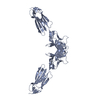
| Title | NMR Structure of ParG symmetric dimer | ||||||
 Components Components | plasmid partition protein ParG | ||||||
 Keywords Keywords | CELL CYCLE / ribbon-helix-helix / dimer / DNA binding | ||||||
| Function / homology |  Function and homology information Function and homology informationDNA-binding transcription repressor activity / core promoter sequence-specific DNA binding / transcription repressor complex / protein-DNA complex / nucleic acid binding / negative regulation of DNA-templated transcription / identical protein binding Similarity search - Function | ||||||
| Biological species |  Salmonella enterica (bacteria) Salmonella enterica (bacteria) | ||||||
| Method | SOLUTION NMR / ARIA protocol (Nilges, M. et al., (1997) J. Mol. Biol. 269, 408-422) was used to deal with ambiguous distance restraints, for some NOE assignments. | ||||||
| Model type details | minimized average | ||||||
 Authors Authors | Golovanov, A.P. / Barilla, D. / Golovanova, M. / Hayes, F. / Lian, L.Y. | ||||||
 Citation Citation |  Journal: Mol.Microbiol. / Year: 2003 Journal: Mol.Microbiol. / Year: 2003Title: ParG, a protein required for active partition of bacterial plasmids, has a dimeric ribbon-helix-helix structure. Authors: Golovanov, A.P. / Barilla, D. / Golovanova, M. / Hayes, F. / Lian, L.Y. #1:  Journal: Mol.Microbiol. / Year: 2000 Journal: Mol.Microbiol. / Year: 2000Title: The partition system of multidrug resistance plasmid TP228 includes a novel protein that epitomizes an evolutionarily distinct subgroup of the ParA superfamily Authors: Hayes, F. | ||||||
| History |
| ||||||
| Remark 650 | HELIX DETERMINATION METHOD: AUTHOR | ||||||
| Remark 700 | SHEET DETERMINATION METHOD: AUTHOR |
- Structure visualization
| Structure viewer | Molecule:  Molmil Molmil Jmol/JSmol Jmol/JSmol |
|---|
- Downloads & links
- Download
| PDBx/mmCIF format |  1p94.cif.gz 1p94.cif.gz | 590.7 KB | Display |  PDBx/mmCIF format PDBx/mmCIF format |
|---|---|---|---|---|
| PDB format |  pdb1p94.ent.gz pdb1p94.ent.gz | 511.5 KB | Display |  PDB format PDB format |
| PDBx/mmJSON format |  1p94.json.gz 1p94.json.gz | Tree view |  PDBx/mmJSON format PDBx/mmJSON format | |
| Others |  Other downloads Other downloads |
-Validation report
| Arichive directory |  https://data.pdbj.org/pub/pdb/validation_reports/p9/1p94 https://data.pdbj.org/pub/pdb/validation_reports/p9/1p94 ftp://data.pdbj.org/pub/pdb/validation_reports/p9/1p94 ftp://data.pdbj.org/pub/pdb/validation_reports/p9/1p94 | HTTPS FTP |
|---|
-Related structure data
| Similar structure data | |
|---|---|
| Other databases |
|
- Links
Links
- Assembly
| Deposited unit | 
| |||||||||
|---|---|---|---|---|---|---|---|---|---|---|
| 1 |
| |||||||||
| NMR ensembles |
|
- Components
| #1: Protein | Mass: 8649.849 Da / Num. of mol.: 2 Source method: isolated from a genetically manipulated source Source: (gene. exp.)  Salmonella enterica (bacteria) / Gene: parG / Plasmid: pET-22-b(+) / Species (production host): Escherichia coli / Production host: Salmonella enterica (bacteria) / Gene: parG / Plasmid: pET-22-b(+) / Species (production host): Escherichia coli / Production host:  |
|---|
-Experimental details
-Experiment
| Experiment | Method: SOLUTION NMR | ||||||||||||||||
|---|---|---|---|---|---|---|---|---|---|---|---|---|---|---|---|---|---|
| NMR experiment |
| ||||||||||||||||
| NMR details | Text: BEST REPRESENTATIVE CONFORMER (MODEL 1) IN THIS ENSEMBLE WAS OBTAINED BY ENERGY MINIMIZATION OF THE AVERAGE STRUCTURE, CALCULATED FOR MODELS 2-11 |
- Sample preparation
| Details |
| ||||||||||||
|---|---|---|---|---|---|---|---|---|---|---|---|---|---|
| Sample conditions | Ionic strength: 150 mM / pH: 5.5 / Pressure: ambient / Temperature: 293 K | ||||||||||||
| Crystal grow | *PLUS Method: other / Details: NMR |
-NMR measurement
| Radiation | Protocol: SINGLE WAVELENGTH / Monochromatic (M) / Laue (L): M | ||||||||||||||||||||
|---|---|---|---|---|---|---|---|---|---|---|---|---|---|---|---|---|---|---|---|---|---|
| Radiation wavelength | Relative weight: 1 | ||||||||||||||||||||
| NMR spectrometer |
|
- Processing
| NMR software |
| ||||||||||||||||||||||||
|---|---|---|---|---|---|---|---|---|---|---|---|---|---|---|---|---|---|---|---|---|---|---|---|---|---|
| Refinement | Method: ARIA protocol (Nilges, M. et al., (1997) J. Mol. Biol. 269, 408-422) was used to deal with ambiguous distance restraints, for some NOE assignments. Software ordinal: 1 Details: The ParG structure is based on 2230 ambiguous NOE restraints, 82 hydrogen bond restraints, and 144 CSI-based dihedral angle restraints. N-terminal region of ParG (1-32) is unstructured. The ...Details: The ParG structure is based on 2230 ambiguous NOE restraints, 82 hydrogen bond restraints, and 144 CSI-based dihedral angle restraints. N-terminal region of ParG (1-32) is unstructured. The C-terminal region (33-76) is structured. | ||||||||||||||||||||||||
| NMR representative | Selection criteria: minimized average structure | ||||||||||||||||||||||||
| NMR ensemble | Conformer selection criteria: structures with the lowest energy,target function Conformers calculated total number: 20 / Conformers submitted total number: 11 |
 Movie
Movie Controller


 PDBj
PDBj NMRPipe