 Movie
Movie Controller


+ Open data

- Basic information
| Entry | Database: PDB / ID: 6ixv | ||||||
|---|---|---|---|---|---|---|---|
| Title | Crystal structure of SH3BP5-Rab11a | ||||||
 Components Components |
| ||||||
 Keywords Keywords | SIGNALING PROTEIN / Rab11 / GEF / SH3BP5 | ||||||
| Function / homology |  Function and homology information Function and homology information: / regulation of protein localization to centrosome / synaptic vesicle endosomal processing / : / regulation of endocytic recycling / establishment of protein localization to organelle / postsynaptic recycling endosome / positive regulation of mitotic cytokinetic process / establishment of vesicle localization / regulation of cilium assembly ...: / regulation of protein localization to centrosome / synaptic vesicle endosomal processing / : / regulation of endocytic recycling / establishment of protein localization to organelle / postsynaptic recycling endosome / positive regulation of mitotic cytokinetic process / establishment of vesicle localization / regulation of cilium assembly / regulation of protein transport / amyloid-beta clearance by transcytosis / neurotransmitter receptor transport, endosome to postsynaptic membrane / exosomal secretion / vesicle-mediated transport in synapse / presynaptic endosome / VxPx cargo-targeting to cilium / plasma membrane to endosome transport / regulation of vesicle-mediated transport / protein transmembrane transport / astral microtubule organization / RAB geranylgeranylation / myosin V binding / multivesicular body assembly / protein localization to cilium / melanosome transport / Golgi to plasma membrane protein transport / establishment of protein localization to membrane / TBC/RABGAPs / protein localization to cell surface / syntaxin binding / dynein light intermediate chain binding / mitotic metaphase chromosome alignment / exocytosis / protein kinase inhibitor activity / cleavage furrow / positive regulation of epithelial cell migration / mitotic spindle assembly / positive regulation of G2/M transition of mitotic cell cycle / transport vesicle / phagocytic vesicle / multivesicular body / vesicle-mediated transport / Anchoring of the basal body to the plasma membrane / guanyl-nucleotide exchange factor activity / cytoplasmic vesicle membrane / trans-Golgi network membrane / regulation of cytokinesis / small monomeric GTPase / protein localization to plasma membrane / positive regulation of protein localization to plasma membrane / Translocation of SLC2A4 (GLUT4) to the plasma membrane / trans-Golgi network / centriole / SH3 domain binding / recycling endosome / regulation of long-term neuronal synaptic plasticity / Schaffer collateral - CA1 synapse / recycling endosome membrane / spindle pole / neuron projection development / endocytic vesicle membrane / Vasopressin regulates renal water homeostasis via Aquaporins / synaptic vesicle membrane / G protein activity / cytoplasmic vesicle / microtubule binding / vesicle / intracellular signal transduction / nuclear body / Golgi membrane / GTPase activity / centrosome / GTP binding / glutamatergic synapse / Golgi apparatus / signal transduction / protein-containing complex / mitochondrion / extracellular exosome / nucleoplasm / cytoplasm / cytosol Similarity search - Function | ||||||
| Biological species |  Homo sapiens (human) Homo sapiens (human) | ||||||
| Method |  X-RAY DIFFRACTION / SYNCHROTRON / MOLECULAR REPLACEMENT / Resolution: 3.8 Å X-RAY DIFFRACTION / SYNCHROTRON / MOLECULAR REPLACEMENT / Resolution: 3.8 Å | ||||||
 Authors Authors | Goto-Ito, S. / Yamagata, A. / Sato, Y. / Fukai, S. | ||||||
| Funding support |  Japan, 1items Japan, 1items
| ||||||
 Citation Citation | Journal: Life Sci Alliance / Year: 2019 Title: Structural basis of guanine nucleotide exchange for Rab11 by SH3BP5. Authors: Goto-Ito, S. / Morooka, N. / Yamagata, A. / Sato, Y. / Sato, K. / Fukai, S. | ||||||
| History |
|
- Structure visualization
| Structure viewer | Molecule: MolmilJmol/JSmol |
|---|
- Downloads & links
-Download
| PDBx/mmCIF format | 6ixv.cif.gz | 322.7 KB | Display | PDBx/mmCIF format |
|---|---|---|---|---|
| PDB format | pdb6ixv.ent.gz | 261.9 KB | Display | PDB format |
| PDBx/mmJSON format | 6ixv.json.gz | Tree view | PDBx/mmJSON format | |
| Others |  Other downloads Other downloads |
-Validation report
| Arichive directory | https://data.pdbj.org/pub/pdb/validation_reports/ix/6ixvftp://data.pdbj.org/pub/pdb/validation_reports/ix/6ixv | HTTPS FTP |
|---|
-Related structure data


-Links
 PDBj
PDBj


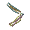
- Assembly
| Deposited unit | 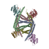
| ||||||||
|---|---|---|---|---|---|---|---|---|---|
| 1 | 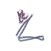
| ||||||||
| 2 | 
| ||||||||
| 3 | 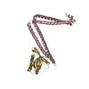
| ||||||||
| 4 | 
| ||||||||
| Unit cell |
|
-Components
| #1: Protein | Mass: 31184.105 Da / Num. of mol.: 4 Source method: isolated from a genetically manipulated source Source: (gene. exp.) Homo sapiens (human) / Gene: SH3BP5, SAB / Production host:  #2: Protein | Mass: 19741.131 Da / Num. of mol.: 4 Source method: isolated from a genetically manipulated source Source: (gene. exp.) Homo sapiens (human) / Gene: RAB11A, RAB11 / Production host: #3: Chemical |   Mass: 94.971 Da / Num. of mol.: 3 / Source method: obtained synthetically / Formula: PO4 Mass: 94.971 Da / Num. of mol.: 3 / Source method: obtained synthetically / Formula: PO4 |
|---|
-Experimental details
-Experiment
| Experiment | Method: X-RAY DIFFRACTION / Number of used crystals: 1 |
|---|
- Sample preparation
| Crystal | Density Matthews: 4.37 Å3/Da / Density % sol: 71.88 % |
|---|---|
| Crystal grow | Temperature: 293 K / Method: vapor diffusion, sitting drop / Details: 0.6M NaCl, 0.1M NaH2PO4 pH 6.8, 16% PEG 2000 |
-Data collection
| Diffraction | Mean temperature: 100 K / Serial crystal experiment: N |
|---|---|
| Diffraction source | Source: SYNCHROTRON / Site: SPring-8 / Beamline: BL41XU / Wavelength: 1 Å |
| Detector | Type: DECTRIS EIGER X 16M / Detector: PIXEL / Date: Jun 2, 2016 |
| Radiation | Protocol: SINGLE WAVELENGTH / Monochromatic (M) / Laue (L): M / Scattering type: x-ray |
| Radiation wavelength | Wavelength: 1 Å / Relative weight: 1 |
| Reflection | Resolution: 3.8→50 Å / Num. obs: 35583 / % possible obs: 100 % / Redundancy: 32.6 % / Rmerge(I) obs: 0.157 / Net I/σ(I): 41.1 |
| Reflection shell | Resolution: 3.8→3.87 Å / Rmerge(I) obs: 1.546 / Num. unique obs: 1505 |
- Processing
| Software |
| ||||||||||||||||||||||||||||||||||||||||||||||||||||||||||||||||||||||||||||||||||||||||||||||||||
|---|---|---|---|---|---|---|---|---|---|---|---|---|---|---|---|---|---|---|---|---|---|---|---|---|---|---|---|---|---|---|---|---|---|---|---|---|---|---|---|---|---|---|---|---|---|---|---|---|---|---|---|---|---|---|---|---|---|---|---|---|---|---|---|---|---|---|---|---|---|---|---|---|---|---|---|---|---|---|---|---|---|---|---|---|---|---|---|---|---|---|---|---|---|---|---|---|---|---|---|
| Refinement | Method to determine structure: MOLECULAR REPLACEMENT / Resolution: 3.8→49.769 Å / SU ML: 0.62 / Cross valid method: FREE R-VALUE / σ(F): 1.34 / Phase error: 31.65 / Stereochemistry target values: ML
| ||||||||||||||||||||||||||||||||||||||||||||||||||||||||||||||||||||||||||||||||||||||||||||||||||
| Solvent computation | Shrinkage radii: 0.9 Å / VDW probe radii: 1.11 Å / Solvent model: FLAT BULK SOLVENT MODEL | ||||||||||||||||||||||||||||||||||||||||||||||||||||||||||||||||||||||||||||||||||||||||||||||||||
| Refinement step | Cycle: LAST / Resolution: 3.8→49.769 Å
| ||||||||||||||||||||||||||||||||||||||||||||||||||||||||||||||||||||||||||||||||||||||||||||||||||
| Refine LS restraints |
| ||||||||||||||||||||||||||||||||||||||||||||||||||||||||||||||||||||||||||||||||||||||||||||||||||
| LS refinement shell |
|
