 Movie
Movie Controller

[English] 日本語
 Yorodumi
- PDB-1p4v: CRYSTAL STRUCTURE OF THE GLYCOSYLASPARAGINASE PRECURSOR D151N MUT... -
+ Open data

- Basic information
| Entry | Database: PDB / ID: 1p4v | ||||||
|---|---|---|---|---|---|---|---|
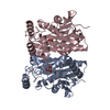
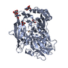
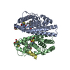
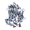
| Title | CRYSTAL STRUCTURE OF THE GLYCOSYLASPARAGINASE PRECURSOR D151N MUTANT WITH GLYCINE | ||||||
 Components Components | N(4)-(Beta-N-acetylglucosaminyl)-L-asparaginase precursor | ||||||
 Keywords Keywords | HYDROLASE / ALPHA BETA / BETA ALPHA / SANDWICH | ||||||
| Function / homology |  Function and homology information Function and homology informationN4-(beta-N-acetylglucosaminyl)-L-asparaginase / N4-(beta-N-acetylglucosaminyl)-L-asparaginase activity / peptidase activity / periplasmic space / proteolysis / cytoplasm Similarity search - Function | ||||||
| Biological species |  Elizabethkingia meningoseptica (bacteria) Elizabethkingia meningoseptica (bacteria) | ||||||
| Method |  X-RAY DIFFRACTION / SYNCHROTRON / MOLECULAR REPLACEMENT / Resolution: 1.9 Å X-RAY DIFFRACTION / SYNCHROTRON / MOLECULAR REPLACEMENT / Resolution: 1.9 Å | ||||||
 Authors Authors | Qian, X. / Guan, C. / Guo, H.C. | ||||||
 Citation Citation | Journal: Structure / Year: 2003 Title: A dual role for an aspartic acid in glycosylasparaginase autoproteolysis. Authors: Qian, X. / Guan, C. / Guo, H.C. | ||||||
| History |
|
- Structure visualization
| Structure viewer | Molecule: MolmilJmol/JSmol |
|---|
- Downloads & links
-Download
| PDBx/mmCIF format | 1p4v.cif.gz | 123.9 KB | Display | PDBx/mmCIF format |
|---|---|---|---|---|
| PDB format | pdb1p4v.ent.gz | 97.2 KB | Display | PDB format |
| PDBx/mmJSON format | 1p4v.json.gz | Tree view | PDBx/mmJSON format | |
| Others |  Other downloads Other downloads |
-Validation report
| Arichive directory | https://data.pdbj.org/pub/pdb/validation_reports/p4/1p4vftp://data.pdbj.org/pub/pdb/validation_reports/p4/1p4v | HTTPS FTP |
|---|
-Related structure data


-Links
 PDBj
PDBj


- Assembly
| Deposited unit | 
| ||||||||
|---|---|---|---|---|---|---|---|---|---|
| 1 |
| ||||||||
| Unit cell |
|
-Components
| #1: Protein | Mass: 32178.645 Da / Num. of mol.: 2 / Fragment: Glycosylasparaginase, alpha and beta chains / Mutation: D151N Source method: isolated from a genetically manipulated source Source: (gene. exp.) Elizabethkingia meningoseptica (bacteria)Plasmid: pMAL-c2x / Production host: References: UniProt: Q47898, N4-(beta-N-acetylglucosaminyl)-L-asparaginase #2: Chemical |   Type: peptide linking / Mass: 75.067 Da / Num. of mol.: 2 / Source method: obtained synthetically / Formula: C2H5NO2 Type: peptide linking / Mass: 75.067 Da / Num. of mol.: 2 / Source method: obtained synthetically / Formula: C2H5NO2#3: Water | ChemComp-HOH / |  Mass: 18.015 Da / Num. of mol.: 212 / Source method: isolated from a natural source / Formula: H2O Mass: 18.015 Da / Num. of mol.: 212 / Source method: isolated from a natural source / Formula: H2O |
|---|
-Experimental details
-Experiment
| Experiment | Method: X-RAY DIFFRACTION / Number of used crystals: 1 |
|---|
- Sample preparation
| Crystal | Density Matthews: 2.13 Å3/Da / Density % sol: 41.89 % | ||||||||||||||||||||||||||||||||||||||||||||||||||||||||||||||||||||||
|---|---|---|---|---|---|---|---|---|---|---|---|---|---|---|---|---|---|---|---|---|---|---|---|---|---|---|---|---|---|---|---|---|---|---|---|---|---|---|---|---|---|---|---|---|---|---|---|---|---|---|---|---|---|---|---|---|---|---|---|---|---|---|---|---|---|---|---|---|---|---|---|
| Crystal grow | Method: evaporation / pH: 7.5 Details: 15% PEG 3300, 0.1M Tris, pH 7.5, 0.2M lithium sulfate, 0.1% sodium azide, 0.05M glycine, EVAPORATION | ||||||||||||||||||||||||||||||||||||||||||||||||||||||||||||||||||||||
| Crystal grow | *PLUS Temperature: 283 K / pH: 7.4 / Method: sparse matrix sampling method / Details: Cui, T., (1999) Acta Crystallogr., D55, 1961. | ||||||||||||||||||||||||||||||||||||||||||||||||||||||||||||||||||||||
| Components of the solutions | *PLUS
|
-Data collection
| Diffraction | Mean temperature: 100 K |
|---|---|
| Diffraction source | Source: SYNCHROTRON / Site: NSLS  / Beamline: X12C / Wavelength: 1.1001 Å / Beamline: X12C / Wavelength: 1.1001 Å |
| Detector | Detector: CCD |
| Radiation | Protocol: SINGLE WAVELENGTH / Monochromatic (M) / Laue (L): M / Scattering type: x-ray |
| Radiation wavelength | Wavelength: 1.1001 Å / Relative weight: 1 |
| Reflection | Resolution: 1.9→20 Å / Num. all: 43152 / Num. obs: 43152 / Rsym value: 0.03 |
| Reflection shell | Resolution: 1.9→1.93 Å / Rsym value: 0.08 |
| Reflection | *PLUS Highest resolution: 1.9 Å / Lowest resolution: 20 Å / Num. obs: 41067 / % possible obs: 95.3 % / Num. measured all: 81010 / Rmerge(I) obs: 0.03 |
| Reflection shell | *PLUS % possible obs: 95.3 % / Rmerge(I) obs: 0.08 |
- Processing
| Software |
| ||||||||||||||||||||
|---|---|---|---|---|---|---|---|---|---|---|---|---|---|---|---|---|---|---|---|---|---|
| Refinement | Method to determine structure: MOLECULAR REPLACEMENT / Resolution: 1.9→20 Å / Cross valid method: THROUGHOUT / σ(F): 0
| ||||||||||||||||||||
| Displacement parameters | Biso mean: 13.5 Å2 | ||||||||||||||||||||
| Refinement step | Cycle: LAST / Resolution: 1.9→20 Å
| ||||||||||||||||||||
| Refine LS restraints |
| ||||||||||||||||||||
| Refinement | *PLUS Highest resolution: 1.9 Å / Lowest resolution: 20 Å / % reflection Rfree: 10 % / Rfactor Rfree: 0.2254 / Rfactor Rwork: 0.1928 | ||||||||||||||||||||
| Solvent computation | *PLUS | ||||||||||||||||||||
| Displacement parameters | *PLUS |
