 Movie
Movie Controller

[English] 日本語
 Yorodumi
- PDB-1p4b: Three-Dimensional Structure Of a Single Chain Fv Fragment Complex... -
+ Open data

- Basic information
| Entry | Database: PDB / ID: 1p4b | ||||||
|---|---|---|---|---|---|---|---|
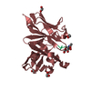
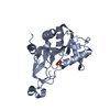
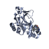
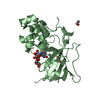
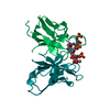
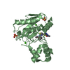
| Title | Three-Dimensional Structure Of a Single Chain Fv Fragment Complexed With The peptide GCN4(7P-14P). | ||||||
 Components Components |
| ||||||
 Keywords Keywords | IMMUNE SYSTEM / Fv antibody / antigen peptide binder / scFv / picomolar binder | ||||||
| Function / homology |  Function and homology information Function and homology informationimmunoglobulin mediated immune response / immunoglobulin complex / antigen binding / adaptive immune response / immune response / extracellular region / plasma membrane Similarity search - Function | ||||||
| Biological species |  | ||||||
| Method |  X-RAY DIFFRACTION / SYNCHROTRON / MOLECULAR REPLACEMENT / Resolution: 2.35 Å X-RAY DIFFRACTION / SYNCHROTRON / MOLECULAR REPLACEMENT / Resolution: 2.35 Å | ||||||
 Authors Authors | Zahnd, C. / Spinelli, S. / Luginbuhl, B. / Jermutus, L. / Amstutz, P. / Cambillau, C. / Pluckthun, A. | ||||||
 Citation Citation | Journal: J.Biol.Chem. / Year: 2004 Title: Directed in Vitro Evolution and Crystallographic Analysis of a Peptide-binding Single Chain Antibody Fragment (scFv) with Low Picomolar Affinity. Authors: Zahnd, C. / Spinelli, S. / Amstutz, P. / Cambillau, C. | ||||||
| History |
| ||||||
| Remark 999 | SEQUENCE an appropriate sequence database reference was not available for chains L, H, and P at the ...SEQUENCE an appropriate sequence database reference was not available for chains L, H, and P at the time of processing. THE AUTHOR STATES THAT THE SEQUENCE HAS NOT BEEN DEPOSITED IN ANY SEQUENCE DATABASE. |
- Structure visualization
| Structure viewer | Molecule: MolmilJmol/JSmol |
|---|
- Downloads & links
-Download
| PDBx/mmCIF format | 1p4b.cif.gz | 60.3 KB | Display | PDBx/mmCIF format |
|---|---|---|---|---|
| PDB format | pdb1p4b.ent.gz | 43.7 KB | Display | PDB format |
| PDBx/mmJSON format | 1p4b.json.gz | Tree view | PDBx/mmJSON format | |
| Others |  Other downloads Other downloads |
-Validation report
| Arichive directory | https://data.pdbj.org/pub/pdb/validation_reports/p4/1p4bftp://data.pdbj.org/pub/pdb/validation_reports/p4/1p4b | HTTPS FTP |
|---|
-Related structure data
| Related structure data |  1p4iC 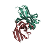 1mfaS S: Starting model for refinement C: citing same article ( |
|---|---|
| Similar structure data |


-Links
 PDBj
PDBj

- Assembly
| Deposited unit | 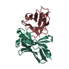
| ||||||||
|---|---|---|---|---|---|---|---|---|---|
| 1 |
| ||||||||
| Unit cell |
|
-Components
| #1: Antibody | Mass: 13333.484 Da / Num. of mol.: 1 Source method: isolated from a genetically manipulated source Source: (gene. exp.)  |
|---|---|
| #2: Antibody | Mass: 13446.813 Da / Num. of mol.: 1 Source method: isolated from a genetically manipulated source Source: (gene. exp.) |
| #3: Protein/peptide | Mass: 1411.648 Da / Num. of mol.: 1 / Source method: obtained synthetically / Details: THE PEPTIDE WAS CHEMICALLY SYNTHESIZED. |
| #4: Water | ChemComp-HOH /  Mass: 18.015 Da / Num. of mol.: 111 / Source method: isolated from a natural source / Formula: H2O Mass: 18.015 Da / Num. of mol.: 111 / Source method: isolated from a natural source / Formula: H2O |
| Has protein modification | Y |
-Experimental details
-Experiment
| Experiment | Method: X-RAY DIFFRACTION / Number of used crystals: 1 |
|---|
- Sample preparation
| Crystal | Density Matthews: 2.02 Å3/Da / Density % sol: 39.22 % |
|---|---|
| Crystal grow | Temperature: 293 K / Method: vapor diffusion, hanging drop / pH: 7.5 Details: PEG 4000, tris/HCl, pH 7.5, VAPOR DIFFUSION, HANGING DROP, temperature 293KK |
-Data collection
| Diffraction | Mean temperature: 100 K |
|---|---|
| Diffraction source | Source: SYNCHROTRON / Site: ESRF  / Beamline: ID14-4 / Wavelength: 0.988 Å / Beamline: ID14-4 / Wavelength: 0.988 Å |
| Detector | Type: ADSC QUANTUM 4 / Detector: CCD |
| Radiation | Protocol: SINGLE WAVELENGTH / Monochromatic (M) / Laue (L): M / Scattering type: x-ray |
| Radiation wavelength | Wavelength: 0.988 Å / Relative weight: 1 |
| Reflection | Resolution: 2.3→20 Å / Num. obs: 10132 / % possible obs: 97 % / Observed criterion σ(F): 2 / Observed criterion σ(I): 2 / Redundancy: 6.7 % / Biso Wilson estimate: 37 Å2 / Rmerge(I) obs: 0.062 / Rsym value: 0.062 / Net I/σ(I): 8.5 |
| Reflection shell | Resolution: 2.3→2.42 Å / Redundancy: 6.9 % / Rmerge(I) obs: 0.171 / Mean I/σ(I) obs: 4.1 / Num. unique all: 1440 / Rsym value: 0.171 / % possible all: 97.2 |
- Processing
| Software |
| |||||||||||||||||||||||||
|---|---|---|---|---|---|---|---|---|---|---|---|---|---|---|---|---|---|---|---|---|---|---|---|---|---|---|
| Refinement | Method to determine structure: MOLECULAR REPLACEMENT Starting model: PDB ENTRY 1MFA Resolution: 2.35→15 Å / σ(F): 2 / σ(I): 2 / Stereochemistry target values: Engh & Huber
| |||||||||||||||||||||||||
| Refinement step | Cycle: LAST / Resolution: 2.35→15 Å
|
