 Movie
Movie Controller

[English] 日本語
 Yorodumi
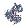
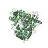
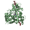
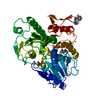
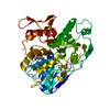
- PDB-1p31: Crystal Structure of UDP-N-acetylmuramic acid:L-alanine Ligase (M... -
+ Open data

- Basic information
| Entry | Database: PDB / ID: 1p31 | ||||||
|---|---|---|---|---|---|---|---|
| Title | Crystal Structure of UDP-N-acetylmuramic acid:L-alanine Ligase (MurC) from Haemophilus influenzae | ||||||
 Components Components | UDP-N-acetylmuramate--alanine ligase | ||||||
 Keywords Keywords | LIGASE / alpha/beta protein | ||||||
| Function / homology |  Function and homology information Function and homology informationUDP-N-acetylmuramate-L-alanine ligase / UDP-N-acetylmuramate-L-alanine ligase activity / peptidoglycan biosynthetic process / cell wall organization / regulation of cell shape / cell division / ATP binding / cytoplasm Similarity search - Function | ||||||
| Biological species |  Haemophilus influenzae (bacteria) Haemophilus influenzae (bacteria) | ||||||
| Method |  X-RAY DIFFRACTION / SYNCHROTRON / SAD / Resolution: 1.85 Å X-RAY DIFFRACTION / SYNCHROTRON / SAD / Resolution: 1.85 Å | ||||||
 Authors Authors | Mol, C.D. / Brooun, A. / Dougan, D.R. / Hilgers, M.T. / Tari, L.W. / Wijnands, R.A. / Knuth, M.W. / McRee, D.E. / Swanson, R.V. | ||||||
 Citation Citation | Journal: J.Bacteriol. / Year: 2003 Title: Crystal Structures of Active Fully Assembled Substrate- and Product-Bound Complexes of UDP-N-Acetylmuramic Acid:L-Alanine Ligase (MurC) from Haemophilus influenzae. Authors: Mol, C.D. / Brooun, A. / Dougan, D.R. / Hilgers, M.T. / Tari, L.W. / Wijnands, R.A. / Knuth, M.W. / McRee, D.E. / Swanson, R.V. | ||||||
| History |
|
- Structure visualization
| Structure viewer | Molecule: MolmilJmol/JSmol |
|---|
- Downloads & links
-Download
| PDBx/mmCIF format | 1p31.cif.gz | 207.4 KB | Display | PDBx/mmCIF format |
|---|---|---|---|---|
| PDB format | pdb1p31.ent.gz | 164.9 KB | Display | PDB format |
| PDBx/mmJSON format | 1p31.json.gz | Tree view | PDBx/mmJSON format | |
| Others |  Other downloads Other downloads |
-Validation report
| Arichive directory | https://data.pdbj.org/pub/pdb/validation_reports/p3/1p31ftp://data.pdbj.org/pub/pdb/validation_reports/p3/1p31 | HTTPS FTP |
|---|
-Related structure data


-Links
 PDBj
PDBj- Assembly
| Deposited unit | 
| ||||||||
|---|---|---|---|---|---|---|---|---|---|
| 1 | 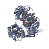
| ||||||||
| 2 | 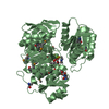
| ||||||||
| Unit cell |
|
-Components
| #1: Protein | Mass: 52760.590 Da / Num. of mol.: 2 Source method: isolated from a genetically manipulated source Source: (gene. exp.) Haemophilus influenzae (bacteria) / Gene: MURC / Plasmid: pSX29 / Production host: References: UniProt: P45066, UDP-N-acetylmuramate-L-alanine ligase #2: Chemical | ChemComp-MG /   Mass: 24.305 Da / Num. of mol.: 4 / Source method: obtained synthetically / Formula: Mg Mass: 24.305 Da / Num. of mol.: 4 / Source method: obtained synthetically / Formula: Mg#3: Chemical | 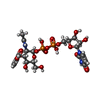  Mass: 677.400 Da / Num. of mol.: 2 / Source method: obtained synthetically / Formula: C20H29N3O19P2 Mass: 677.400 Da / Num. of mol.: 2 / Source method: obtained synthetically / Formula: C20H29N3O19P2#4: Water | ChemComp-HOH / |  Mass: 18.015 Da / Num. of mol.: 700 / Source method: isolated from a natural source / Formula: H2O Mass: 18.015 Da / Num. of mol.: 700 / Source method: isolated from a natural source / Formula: H2OHas protein modification | Y | |
|---|
-Experimental details
-Experiment
| Experiment | Method: X-RAY DIFFRACTION / Number of used crystals: 1 |
|---|
- Sample preparation
| Crystal | Density Matthews: 2.36 Å3/Da / Density % sol: 47.96 % | ||||||||||||||||||||||||||||||||||||||||||||||||||||||||||||||||||||||
|---|---|---|---|---|---|---|---|---|---|---|---|---|---|---|---|---|---|---|---|---|---|---|---|---|---|---|---|---|---|---|---|---|---|---|---|---|---|---|---|---|---|---|---|---|---|---|---|---|---|---|---|---|---|---|---|---|---|---|---|---|---|---|---|---|---|---|---|---|---|---|---|
| Crystal grow | Temperature: 277 K / Method: nanovolume / pH: 8.1 Details: PEG 4000, glycerol, magnesium chloride, Tris, pH 8.1, nanovolume, temperature 277K | ||||||||||||||||||||||||||||||||||||||||||||||||||||||||||||||||||||||
| Crystal grow | *PLUS Temperature: 4 ℃ / pH: 7.9 / Method: vapor diffusion, sitting drop | ||||||||||||||||||||||||||||||||||||||||||||||||||||||||||||||||||||||
| Components of the solutions | *PLUS
|
-Data collection
| Diffraction | Mean temperature: 100 K |
|---|---|
| Diffraction source | Source: SYNCHROTRON / Site: ALS  / Beamline: 5.0.3 / Wavelength: 1 Å / Beamline: 5.0.3 / Wavelength: 1 Å |
| Detector | Type: ADSC QUANTUM 4 / Detector: CCD / Date: Jul 31, 2002 |
| Radiation | Monochromator: Si crystal / Protocol: SINGLE WAVELENGTH / Monochromatic (M) / Laue (L): M / Scattering type: x-ray |
| Radiation wavelength | Wavelength: 1 Å / Relative weight: 1 |
| Reflection | Resolution: 1.85→25 Å / Num. all: 86600 / Num. obs: 77944 / % possible obs: 90 % / Observed criterion σ(F): 2 / Observed criterion σ(I): 2 / Redundancy: 4.45 % / Biso Wilson estimate: 13.3 Å2 / Rmerge(I) obs: 0.064 / Net I/σ(I): 10.8 |
| Reflection shell | Resolution: 1.85→1.89 Å / Redundancy: 4.5 % / Rmerge(I) obs: 0.45 / Mean I/σ(I) obs: 3 / Num. unique all: 5314 / % possible all: 93 |
| Reflection | *PLUS % possible obs: 90 % / Num. measured all: 347154 |
| Reflection shell | *PLUS % possible obs: 93 % |
- Processing
| Software |
| |||||||||||||||||||||||||||||||||||||||||||||||||||||||||||||||||||||||||||||||||||||||||||||||||||||||||||||||||||||||||||||||||||||||||||||||||||||||||||||||||||||||||||||||
|---|---|---|---|---|---|---|---|---|---|---|---|---|---|---|---|---|---|---|---|---|---|---|---|---|---|---|---|---|---|---|---|---|---|---|---|---|---|---|---|---|---|---|---|---|---|---|---|---|---|---|---|---|---|---|---|---|---|---|---|---|---|---|---|---|---|---|---|---|---|---|---|---|---|---|---|---|---|---|---|---|---|---|---|---|---|---|---|---|---|---|---|---|---|---|---|---|---|---|---|---|---|---|---|---|---|---|---|---|---|---|---|---|---|---|---|---|---|---|---|---|---|---|---|---|---|---|---|---|---|---|---|---|---|---|---|---|---|---|---|---|---|---|---|---|---|---|---|---|---|---|---|---|---|---|---|---|---|---|---|---|---|---|---|---|---|---|---|---|---|---|---|---|---|---|---|---|
| Refinement | Method to determine structure: SAD / Resolution: 1.85→25 Å / Cor.coef. Fo:Fc: 0.957 / Cor.coef. Fo:Fc free: 0.936 / SU B: 2.59 / SU ML: 0.08 / TLS residual ADP flag: LIKELY RESIDUAL / Isotropic thermal model: isotropic / Cross valid method: THROUGHOUT / σ(F): 0 / σ(I): 0 / ESU R: 0.144 / ESU R Free: 0.134 / Stereochemistry target values: MAXIMUM LIKELIHOOD
| |||||||||||||||||||||||||||||||||||||||||||||||||||||||||||||||||||||||||||||||||||||||||||||||||||||||||||||||||||||||||||||||||||||||||||||||||||||||||||||||||||||||||||||||
| Solvent computation | Ion probe radii: 0.8 Å / Shrinkage radii: 0.8 Å / VDW probe radii: 1.4 Å / Solvent model: BABINET MODEL WITH MASK | |||||||||||||||||||||||||||||||||||||||||||||||||||||||||||||||||||||||||||||||||||||||||||||||||||||||||||||||||||||||||||||||||||||||||||||||||||||||||||||||||||||||||||||||
| Displacement parameters | Biso mean: 13.859 Å2
| |||||||||||||||||||||||||||||||||||||||||||||||||||||||||||||||||||||||||||||||||||||||||||||||||||||||||||||||||||||||||||||||||||||||||||||||||||||||||||||||||||||||||||||||
| Refinement step | Cycle: LAST / Resolution: 1.85→25 Å
| |||||||||||||||||||||||||||||||||||||||||||||||||||||||||||||||||||||||||||||||||||||||||||||||||||||||||||||||||||||||||||||||||||||||||||||||||||||||||||||||||||||||||||||||
| Refine LS restraints |
| |||||||||||||||||||||||||||||||||||||||||||||||||||||||||||||||||||||||||||||||||||||||||||||||||||||||||||||||||||||||||||||||||||||||||||||||||||||||||||||||||||||||||||||||
| LS refinement shell | Resolution: 1.85→1.898 Å / Total num. of bins used: 20 /
| |||||||||||||||||||||||||||||||||||||||||||||||||||||||||||||||||||||||||||||||||||||||||||||||||||||||||||||||||||||||||||||||||||||||||||||||||||||||||||||||||||||||||||||||
| Refinement TLS params. | Method: refined / Refine-ID: X-RAY DIFFRACTION
| |||||||||||||||||||||||||||||||||||||||||||||||||||||||||||||||||||||||||||||||||||||||||||||||||||||||||||||||||||||||||||||||||||||||||||||||||||||||||||||||||||||||||||||||
| Refinement TLS group |
| |||||||||||||||||||||||||||||||||||||||||||||||||||||||||||||||||||||||||||||||||||||||||||||||||||||||||||||||||||||||||||||||||||||||||||||||||||||||||||||||||||||||||||||||
| Refinement | *PLUS Lowest resolution: 25 Å / Rfactor Rfree: 0.208 / Rfactor Rwork: 0.169 | |||||||||||||||||||||||||||||||||||||||||||||||||||||||||||||||||||||||||||||||||||||||||||||||||||||||||||||||||||||||||||||||||||||||||||||||||||||||||||||||||||||||||||||||
| Solvent computation | *PLUS | |||||||||||||||||||||||||||||||||||||||||||||||||||||||||||||||||||||||||||||||||||||||||||||||||||||||||||||||||||||||||||||||||||||||||||||||||||||||||||||||||||||||||||||||
| Displacement parameters | *PLUS | |||||||||||||||||||||||||||||||||||||||||||||||||||||||||||||||||||||||||||||||||||||||||||||||||||||||||||||||||||||||||||||||||||||||||||||||||||||||||||||||||||||||||||||||
| Refine LS restraints | *PLUS
| |||||||||||||||||||||||||||||||||||||||||||||||||||||||||||||||||||||||||||||||||||||||||||||||||||||||||||||||||||||||||||||||||||||||||||||||||||||||||||||||||||||||||||||||
| LS refinement shell | *PLUS Highest resolution: 1.85 Å / Lowest resolution: 1.9 Å |
