 Movie
Movie Controller


+ Open data

- Basic information
| Entry | Database: PDB / ID: 1p1h | ||||||
|---|---|---|---|---|---|---|---|
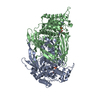
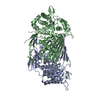
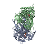
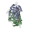
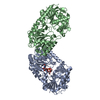
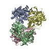
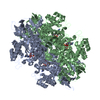
| Title | Crystal structure of the 1L-myo-inositol/NAD+ complex | ||||||
 Components Components | Inositol-3-phosphate synthase | ||||||
 Keywords Keywords | ISOMERASE / NAD+ / 1L-myo-inositol 1-phosphate | ||||||
| Function / homology |  Function and homology information Function and homology informationSynthesis of IP2, IP, and Ins in the cytosol / inositol-3-phosphate synthase / inositol-3-phosphate synthase activity / inositol biosynthetic process / phospholipid biosynthetic process / cytoplasm Similarity search - Function | ||||||
| Biological species |  | ||||||
| Method |  X-RAY DIFFRACTION / SYNCHROTRON / MOLECULAR REPLACEMENT / Resolution: 1.95 Å X-RAY DIFFRACTION / SYNCHROTRON / MOLECULAR REPLACEMENT / Resolution: 1.95 Å | ||||||
 Authors Authors | Jin, X. / Geiger, J.H. | ||||||
 Citation Citation | Journal: Acta Crystallogr.,Sect.D / Year: 2003 Title: Structures of NAD(+)- and NADH-bound 1-l-myo-inositol 1-phosphate synthase. Authors: Jin, X. / Geiger, J.H. | ||||||
| History |
| ||||||
| Remark 999 | The differences between the authors' sequence and the database reference sequence are known ... The differences between the authors' sequence and the database reference sequence are known conflicts and have been documented in Swiss Prot entry P11986. |
- Structure visualization
| Structure viewer | Molecule: MolmilJmol/JSmol |
|---|
- Downloads & links
-Download
| PDBx/mmCIF format | 1p1h.cif.gz | 434.1 KB | Display | PDBx/mmCIF format |
|---|---|---|---|---|
| PDB format | pdb1p1h.ent.gz | 353.4 KB | Display | PDB format |
| PDBx/mmJSON format | 1p1h.json.gz | Tree view | PDBx/mmJSON format | |
| Others |  Other downloads Other downloads |
-Validation report
| Arichive directory | https://data.pdbj.org/pub/pdb/validation_reports/p1/1p1hftp://data.pdbj.org/pub/pdb/validation_reports/p1/1p1h | HTTPS FTP |
|---|
-Related structure data


-Links
 PDBj
PDBj- Assembly
| Deposited unit | 
| ||||||||
|---|---|---|---|---|---|---|---|---|---|
| 1 |
| ||||||||
| Unit cell |
| ||||||||
| Details | The biological assembly is a tetramer in the asymmetric unit. |
-Components
| #1: Protein | Mass: 59707.766 Da / Num. of mol.: 4 Source method: isolated from a genetically manipulated source Source: (gene. exp.) Production host:  #2: Chemical | ChemComp-NAD /   Mass: 663.425 Da / Num. of mol.: 4 / Source method: obtained synthetically / Formula: C21H27N7O14P2 / Comment: NAD*YM Mass: 663.425 Da / Num. of mol.: 4 / Source method: obtained synthetically / Formula: C21H27N7O14P2 / Comment: NAD*YM#3: Water | ChemComp-HOH / |  Mass: 18.015 Da / Num. of mol.: 1529 / Source method: isolated from a natural source / Formula: H2O Mass: 18.015 Da / Num. of mol.: 1529 / Source method: isolated from a natural source / Formula: H2O |
|---|
-Experimental details
-Experiment
| Experiment | Method: X-RAY DIFFRACTION / Number of used crystals: 1 |
|---|
- Sample preparation
| Crystal | Density Matthews: 3.01 Å3/Da / Density % sol: 59.16 % | ||||||||||||||||||||||||||||||||||||||||||
|---|---|---|---|---|---|---|---|---|---|---|---|---|---|---|---|---|---|---|---|---|---|---|---|---|---|---|---|---|---|---|---|---|---|---|---|---|---|---|---|---|---|---|---|
| Crystal grow | Temperature: 298 K / Method: vapor diffusion, hanging drop / pH: 4.6 Details: PEG 8000, sodium acetate, pH 4.6, VAPOR DIFFUSION, HANGING DROP, temperature 298K | ||||||||||||||||||||||||||||||||||||||||||
| Crystal grow | *PLUS pH: 4.5 / Method: vapor diffusion, hanging dropDetails: J., Stein. A., (2000) Acta Crystallogr., Sect.D, 56, 348. | ||||||||||||||||||||||||||||||||||||||||||
| Components of the solutions | *PLUS
|
-Data collection
| Diffraction | Mean temperature: 100 K |
|---|---|
| Diffraction source | Source: SYNCHROTRON / Site: APS  / Beamline: 14-BM-D / Wavelength: 0.9 Å / Beamline: 14-BM-D / Wavelength: 0.9 Å |
| Detector | Type: ADSC QUANTUM 4 / Detector: CCD / Date: Apr 8, 2002 |
| Radiation | Protocol: SINGLE WAVELENGTH / Monochromatic (M) / Laue (L): M / Scattering type: x-ray |
| Radiation wavelength | Wavelength: 0.9 Å / Relative weight: 1 |
| Reflection | Resolution: 1.95→50 Å / Num. obs: 203310 / % possible obs: 96.6 % / Observed criterion σ(F): 2 / Observed criterion σ(I): 2 |
| Reflection shell | Resolution: 1.9→1.97 Å / % possible all: 90.4 |
| Reflection | *PLUS Num. measured all: 214444 / Rmerge(I) obs: 0.064 |
| Reflection shell | *PLUS % possible obs: 99.2 % / Rmerge(I) obs: 0.422 / Mean I/σ(I) obs: 2.5 |
- Processing
| Software |
| ||||||||||||||||||||
|---|---|---|---|---|---|---|---|---|---|---|---|---|---|---|---|---|---|---|---|---|---|
| Refinement | Method to determine structure: MOLECULAR REPLACEMENT / Resolution: 1.95→10 Å / Cross valid method: THROUGHOUT / σ(F): 2 / Stereochemistry target values: Engh & Huber
| ||||||||||||||||||||
| Displacement parameters |
| ||||||||||||||||||||
| Refinement step | Cycle: LAST / Resolution: 1.95→10 Å
| ||||||||||||||||||||
| Refine LS restraints |
| ||||||||||||||||||||
| Xplor file |
| ||||||||||||||||||||
| Refinement | *PLUS % reflection Rfree: 10 % | ||||||||||||||||||||
| Solvent computation | *PLUS | ||||||||||||||||||||
| Displacement parameters | *PLUS | ||||||||||||||||||||
| Refine LS restraints | *PLUS
|
