 Movie
Movie Controller

[English] 日本語
 Yorodumi
- PDB-1jki: myo-Inositol-1-phosphate Synthase Complexed with an Inhibitor, 2-... -
+ Open data

- Basic information
| Entry | Database: PDB / ID: 1jki | ||||||
|---|---|---|---|---|---|---|---|
| Title | myo-Inositol-1-phosphate Synthase Complexed with an Inhibitor, 2-deoxy-glucitol-6-phosphate | ||||||
 Components Components | myo-inositol-1-phosphate synthase | ||||||
 Keywords Keywords | ISOMERASE / Rossmann fold / inhibitor-bound structure | ||||||
| Function / homology |  Function and homology information Function and homology informationSynthesis of IP2, IP, and Ins in the cytosol / inositol-3-phosphate synthase / inositol-3-phosphate synthase activity / inositol biosynthetic process / phospholipid biosynthetic process / cytoplasm Similarity search - Function | ||||||
| Biological species |  | ||||||
| Method |  X-RAY DIFFRACTION / SYNCHROTRON / MOLECULAR REPLACEMENT / Resolution: 2.2 Å X-RAY DIFFRACTION / SYNCHROTRON / MOLECULAR REPLACEMENT / Resolution: 2.2 Å | ||||||
 Authors Authors | Stein, A.J. / Geiger, J.H. | ||||||
 Citation Citation | Journal: J.Biol.Chem. / Year: 2002 Title: The crystal structure and mechanism of 1-L-myo-inositol- 1-phosphate synthase Authors: Stein, A.J. / Geiger, J.H. #1: Journal: Acta Crystallogr.,Sect.D / Year: 2000Title: MIP Synthase Structural Studies Authors: Stein, A.J. / Geiger, J.H. | ||||||
| History |
|
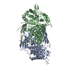
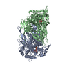
- Structure visualization
| Structure viewer | Molecule: MolmilJmol/JSmol |
|---|
- Downloads & links
-Download
| PDBx/mmCIF format | 1jki.cif.gz | 235.3 KB | Display | PDBx/mmCIF format |
|---|---|---|---|---|
| PDB format | pdb1jki.ent.gz | 185.9 KB | Display | PDB format |
| PDBx/mmJSON format | 1jki.json.gz | Tree view | PDBx/mmJSON format | |
| Others |  Other downloads Other downloads |
-Validation report
| Arichive directory | https://data.pdbj.org/pub/pdb/validation_reports/jk/1jkiftp://data.pdbj.org/pub/pdb/validation_reports/jk/1jki | HTTPS FTP |
|---|
-Related structure data
| Related structure data | 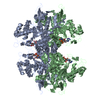 1jkfSC S: Starting model for refinement C: citing same article ( |
|---|---|
| Similar structure data |


-Links
 PDBj
PDBj


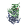
- Assembly
| Deposited unit | 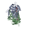
| ||||||||
|---|---|---|---|---|---|---|---|---|---|
| 1 | 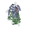
| ||||||||
| Unit cell |
| ||||||||
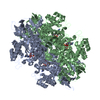
| Details | The biological assembly is a tetramer . The tetramer has 222 symmetry with a non-crystallographic 2-fold axis relating the two monomers in the assymmetric unit and a crystallographic 2-fold axis relating the two dimers that make up the tetramer. The operations are x, y, z and -x, y, -z. |
-Components
| #1: Protein | Mass: 59707.766 Da / Num. of mol.: 2 Source method: isolated from a genetically manipulated source Source: (gene. exp.) Production host:  #2: Chemical |   Mass: 18.038 Da / Num. of mol.: 2 / Source method: obtained synthetically / Formula: H4N Mass: 18.038 Da / Num. of mol.: 2 / Source method: obtained synthetically / Formula: H4N#3: Chemical | 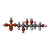  Mass: 246.152 Da / Num. of mol.: 2 / Source method: obtained synthetically / Formula: C6H15O8P Mass: 246.152 Da / Num. of mol.: 2 / Source method: obtained synthetically / Formula: C6H15O8P#4: Chemical |   Mass: 665.441 Da / Num. of mol.: 2 / Source method: obtained synthetically / Formula: C21H29N7O14P2 Mass: 665.441 Da / Num. of mol.: 2 / Source method: obtained synthetically / Formula: C21H29N7O14P2#5: Water | ChemComp-HOH / |  Mass: 18.015 Da / Num. of mol.: 618 / Source method: isolated from a natural source / Formula: H2O Mass: 18.015 Da / Num. of mol.: 618 / Source method: isolated from a natural source / Formula: H2O |
|---|
-Experimental details
-Experiment
| Experiment | Method: X-RAY DIFFRACTION / Number of used crystals: 1 |
|---|
- Sample preparation
| Crystal | Density Matthews: 3.09 Å3/Da / Density % sol: 60.24 % | ||||||||||||||||||||||||||||||||||||||||||
|---|---|---|---|---|---|---|---|---|---|---|---|---|---|---|---|---|---|---|---|---|---|---|---|---|---|---|---|---|---|---|---|---|---|---|---|---|---|---|---|---|---|---|---|
| Crystal grow | Temperature: 298 K / Method: co-crystalliztion / pH: 4.5 Details: PEG 8000, sodium acetate, NAD, and 2-deoxy-glucitol-6-phosphate, pH 4.5, co-crystalliztion, temperature 298K | ||||||||||||||||||||||||||||||||||||||||||
| Crystal grow | *PLUS Method: vapor diffusion, hanging dropDetails: J., Stein. A., (2000) Acta Crystallogr., Sect.D, 56, 348. | ||||||||||||||||||||||||||||||||||||||||||
| Components of the solutions | *PLUS
|
-Data collection
| Diffraction | Mean temperature: 123 K |
|---|---|
| Diffraction source | Source: SYNCHROTRON / Site: APS  / Beamline: 19-ID / Wavelength: 0.97942 Å / Beamline: 19-ID / Wavelength: 0.97942 Å |
| Detector | Type: SBC / Detector: CCD / Date: Mar 20, 2001 |
| Radiation | Protocol: SINGLE WAVELENGTH / Monochromatic (M) / Laue (L): M / Scattering type: x-ray |
| Radiation wavelength | Wavelength: 0.97942 Å / Relative weight: 1 |
| Reflection | Resolution: 2.2→50 Å / Num. all: 92832 / Num. obs: 76517 / % possible obs: 89.2 % / Observed criterion σ(F): 1 / Observed criterion σ(I): 1 / Rsym value: 0.082 / Net I/σ(I): 13.9 |
| Reflection shell | Resolution: 2.2→2.3 Å / Mean I/σ(I) obs: 4.6 / Rsym value: 0.304 / % possible all: 89.2 |
| Reflection | *PLUS % possible obs: 94.9 % / Rmerge(I) obs: 0.082 |
| Reflection shell | *PLUS % possible obs: 96.3 % / Rmerge(I) obs: 0.304 |
- Processing
| Software |
| |||||||||||||||||||||||||
|---|---|---|---|---|---|---|---|---|---|---|---|---|---|---|---|---|---|---|---|---|---|---|---|---|---|---|
| Refinement | Method to determine structure: MOLECULAR REPLACEMENT Starting model: MIP synthase, PDB 1JKF Resolution: 2.2→10 Å / σ(F): 2 / Stereochemistry target values: CNS, Terwilliger
| |||||||||||||||||||||||||
| Refinement step | Cycle: LAST / Resolution: 2.2→10 Å
| |||||||||||||||||||||||||
| Refine LS restraints |
| |||||||||||||||||||||||||
| LS refinement shell | Resolution: 2.2→2.3 Å /
| |||||||||||||||||||||||||
| Refinement | *PLUS % reflection Rfree: 10 % / Rfactor all: 0.285 / Rfactor obs: 0.208 / Rfactor Rfree: 0.276 / Rfactor Rwork: 0.208 | |||||||||||||||||||||||||
| Solvent computation | *PLUS | |||||||||||||||||||||||||
| Displacement parameters | *PLUS | |||||||||||||||||||||||||
| LS refinement shell | *PLUS Rfactor Rfree: 0.365 / Rfactor Rwork: 0.269 |
