 Movie
Movie Controller

[English] 日本語
 Yorodumi
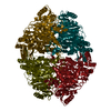
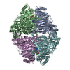
- PDB-1ovm: Crystal structure of Indolepyruvate decarboxylase from Enterobact... -
+ Open data

- Basic information
| Entry | Database: PDB / ID: 1ovm | ||||||
|---|---|---|---|---|---|---|---|
| Title | Crystal structure of Indolepyruvate decarboxylase from Enterobacter cloacae | ||||||
 Components Components | Indole-3-pyruvate decarboxylase | ||||||
 Keywords Keywords | LYASE / thiamine diphosphate / indole-3-acetic acid / indolepyruvate / tdp dependent enzyme / decarboxylase | ||||||
| Function / homology |  Function and homology information Function and homology informationindolepyruvate decarboxylase / indolepyruvate decarboxylase activity / auxin biosynthetic process / : / pyruvate decarboxylase activity / thiamine pyrophosphate binding / magnesium ion binding / cytosol Similarity search - Function | ||||||
| Biological species |  Enterobacter cloacae (bacteria) Enterobacter cloacae (bacteria) | ||||||
| Method |  X-RAY DIFFRACTION / SYNCHROTRON / MOLECULAR REPLACEMENT / Resolution: 2.65 Å X-RAY DIFFRACTION / SYNCHROTRON / MOLECULAR REPLACEMENT / Resolution: 2.65 Å | ||||||
 Authors Authors | Schutz, A. / Sandalova, T. / Ricagno, S. / Hubner, G. / Konig, S. / Schneider, G. | ||||||
 Citation Citation | Journal: Eur.J.Biochem. / Year: 2003 Title: Crystal structure of thiamindiphosphate-dependent indolepyruvate decarboxylase from Enterobacter cloacae, an enzyme involved in the biosynthesis of the plant hormone indole-3-acetic acid Authors: Schutz, A. / Sandalova, T. / Ricagno, S. / Hubner, G. / Konig, S. / Schneider, G. | ||||||
| History |
|
- Structure visualization
| Structure viewer | Molecule: MolmilJmol/JSmol |
|---|
- Downloads & links
-Download
| PDBx/mmCIF format | 1ovm.cif.gz | 413.7 KB | Display | PDBx/mmCIF format |
|---|---|---|---|---|
| PDB format | pdb1ovm.ent.gz | 337.9 KB | Display | PDB format |
| PDBx/mmJSON format | 1ovm.json.gz | Tree view | PDBx/mmJSON format | |
| Others |  Other downloads Other downloads |
-Validation report
| Arichive directory | https://data.pdbj.org/pub/pdb/validation_reports/ov/1ovmftp://data.pdbj.org/pub/pdb/validation_reports/ov/1ovm | HTTPS FTP |
|---|
-Related structure data
| Related structure data | 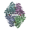 1zpdS S: Starting model for refinement |
|---|---|
| Similar structure data |


-Links
 PDBj
PDBj


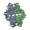
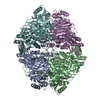
- Assembly
| Deposited unit | 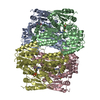
| ||||||||
|---|---|---|---|---|---|---|---|---|---|
| 1 |
| ||||||||
| Unit cell |
|
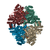
-Components
| #1: Protein | Mass: 60087.289 Da / Num. of mol.: 4 Source method: isolated from a genetically manipulated source Source: (gene. exp.) Enterobacter cloacae (bacteria) / Gene: IPDC / Plasmid: pIP362 / Production host: #2: Chemical | ChemComp-MG /   Mass: 24.305 Da / Num. of mol.: 4 / Source method: obtained synthetically / Formula: Mg Mass: 24.305 Da / Num. of mol.: 4 / Source method: obtained synthetically / Formula: Mg#3: Chemical | ChemComp-TPP /   Mass: 425.314 Da / Num. of mol.: 4 / Source method: obtained synthetically / Formula: C12H19N4O7P2S Mass: 425.314 Da / Num. of mol.: 4 / Source method: obtained synthetically / Formula: C12H19N4O7P2S#4: Water | ChemComp-HOH / |  Mass: 18.015 Da / Num. of mol.: 347 / Source method: isolated from a natural source / Formula: H2O Mass: 18.015 Da / Num. of mol.: 347 / Source method: isolated from a natural source / Formula: H2O |
|---|
-Experimental details
-Experiment
| Experiment | Method: X-RAY DIFFRACTION / Number of used crystals: 1 |
|---|
- Sample preparation
| Crystal | Density Matthews: 2.28 Å3/Da / Density % sol: 45.71 % | ||||||||||||||||||||||||||||||||||||
|---|---|---|---|---|---|---|---|---|---|---|---|---|---|---|---|---|---|---|---|---|---|---|---|---|---|---|---|---|---|---|---|---|---|---|---|---|---|
| Crystal grow | Temperature: 293 K / Method: vapor diffusion, hanging drop / pH: 5 Details: PEGmme, Na citrate, pH 5.0, VAPOR DIFFUSION, HANGING DROP, temperature 293K | ||||||||||||||||||||||||||||||||||||
| Crystal grow | *PLUS Temperature: 20 ℃ / Method: vapor diffusion, hanging drop | ||||||||||||||||||||||||||||||||||||
| Components of the solutions | *PLUS
|
-Data collection
| Diffraction | Mean temperature: 110 K |
|---|---|
| Diffraction source | Source: SYNCHROTRON / Site: ESRF  / Beamline: ID29 / Wavelength: 0.979 Å / Beamline: ID29 / Wavelength: 0.979 Å |
| Detector | Type: ADSC QUANTUM 4 / Detector: CCD / Date: Apr 11, 2002 |
| Radiation | Protocol: SINGLE WAVELENGTH / Monochromatic (M) / Laue (L): M / Scattering type: x-ray |
| Radiation wavelength | Wavelength: 0.979 Å / Relative weight: 1 |
| Reflection | Resolution: 2.65→29.56 Å / Num. all: 63376 / Num. obs: 63376 / % possible obs: 99.9 % / Observed criterion σ(F): 0 / Observed criterion σ(I): 0 / Biso Wilson estimate: 44.9 Å2 / Limit h max: 49 / Limit h min: 0 / Limit k max: 57 / Limit k min: 0 / Limit l max: 40 / Limit l min: 0 / Observed criterion F max: 2754137.27 / Observed criterion F min: 25.6 / Rsym value: 0.087 / Net I/σ(I): 7.8 |
| Reflection shell | Resolution: 2.65→2.77 Å / Mean I/σ(I) obs: 2 / Rsym value: 0.369 / % possible all: 99.9 |
| Reflection | *PLUS Num. obs: 63426 / Num. measured all: 315465 / Rmerge(I) obs: 0.087 |
| Reflection shell | *PLUS % possible obs: 99.9 % / Rmerge(I) obs: 0.369 |
- Processing
| Software |
| |||||||||||||||||||||||||
|---|---|---|---|---|---|---|---|---|---|---|---|---|---|---|---|---|---|---|---|---|---|---|---|---|---|---|
| Refinement | Method to determine structure: MOLECULAR REPLACEMENT Starting model: PDB ENTRY 1ZPD Resolution: 2.65→29.56 Å / Rfactor Rfree error: 0.004 / Occupancy max: 1 / Occupancy min: 1 / Cross valid method: THROUGHOUT / σ(F): 0 / Stereochemistry target values: MAXIMUM LIKELIHOOD
| |||||||||||||||||||||||||
| Solvent computation | Solvent model: CNS bulk solvent model used / Bsol: 22.125 Å2 / ksol: 0.33713 e/Å3 | |||||||||||||||||||||||||
| Displacement parameters | Biso max: 93.33 Å2 / Biso mean: 33.42 Å2 / Biso min: 2.47 Å2
| |||||||||||||||||||||||||
| Refine analyze |
| |||||||||||||||||||||||||
| Refinement step | Cycle: LAST / Resolution: 2.65→29.56 Å
| |||||||||||||||||||||||||
| Refine LS restraints |
| |||||||||||||||||||||||||
| LS refinement shell | Resolution: 2.65→2.77 Å / Rfactor Rfree error: 0.014
| |||||||||||||||||||||||||
| Xplor file |
| |||||||||||||||||||||||||
| Software | *PLUS Name: CNS / Classification: refinement | |||||||||||||||||||||||||
| Refinement | *PLUS | |||||||||||||||||||||||||
| Solvent computation | *PLUS | |||||||||||||||||||||||||
| Displacement parameters | *PLUS | |||||||||||||||||||||||||
| Refine LS restraints | *PLUS
|
